2EVE | pdb_00002eve

X-Ray Crystal Structure of Protein PSPTO5229 from Pseudomonas syringae. Northeast Structural Genomics Consortium Target PsR62


Experimental Data Snapshot

  • Method: X-RAY DIFFRACTION
  • Resolution: 1.60 Å
  • R-Value Free: 
    0.217 (Depositor), 0.232 (DCC) 
  • R-Value Work: 
    0.191 (Depositor), 0.202 (DCC) 
  • R-Value Observed: 
    0.191 (Depositor) 

wwPDB Validation 3D Report Full Report

Validation slider image for 2EVE

Ligand Structure Quality Assessment 


This is version 1.4 of the entry. See complete history

Literature

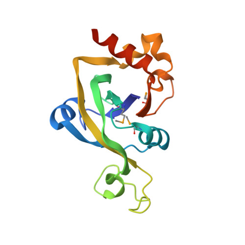
Structural genomics reveals EVE as a new ASCH/PUA-related domain.

Bertonati, C.Punta, M.Fischer, M.Yachdav, G.Forouhar, F.Zhou, W.Kuzin, A.P.Seetharaman, J.Abashidze, M.Ramelot, T.A.Kennedy, M.A.Cort, J.R.Belachew, A.Hunt, J.F.Tong, L.Montelione, G.T.Rost, B.

(2009) Proteins 75: 760-773

  • DOI: https://doi.org/10.1002/prot.22287
  • Primary Citation Related Structures: 
    1ZCE, 2EVE, 2G2X, 2GBS

  • PubMed Abstract: 

    We report on several proteins recently solved by structural genomics consortia, in particular by the Northeast Structural Genomics consortium (NESG). The proteins considered in this study differ substantially in their sequences but they share a similar structural core, characterized by a pseudobarrel five-stranded beta sheet. This core corresponds to the PUA domain-like architecture in the SCOP database. By connecting sequence information with structural knowledge, we characterize a new subgroup of these proteins that we propose to be distinctly different from previously described PUA domain-like domains such as PUA proper or ASCH. We refer to these newly defined domains as EVE. Although EVE may have retained the ability of PUA domains to bind RNA, the available experimental and computational data suggests that both the details of its molecular function and its cellular function differ from those of other PUA domain-like domains. This study of EVE and its relatives illustrates how the combination of structure and genomics creates new insights by connecting a cornucopia of structures that map to the same evolutionary potential. Primary sequence information alone would have not been sufficient to reveal these evolutionary links.


  • Organizational Affiliation
    • Department of Biochemistry and Molecular Biophysics, Columbia University, New York, NY 10032, USA. claudia.bertonati@uniroma1.it

Macromolecule Content 

  • Total Structure Weight: 18.5 kDa 
  • Atom Count: 1,430 
  • Modeled Residue Count: 149 
  • Deposited Residue Count: 157 
  • Unique protein chains: 1

Macromolecules

Find similar proteins by:|  3D Structure
Entity ID: 1
MoleculeChains  Sequence LengthOrganismDetailsImage
hypothetical protein PSPTO5229157Pseudomonas syringae pv. tomato str. DC3000Mutation(s): 4 
Gene Names: 1186914
UniProt
Find proteins for Q87UR7 (Pseudomonas syringae pv. tomato (strain ATCC BAA-871 / DC3000))
Explore Q87UR7 
Go to UniProtKB:  Q87UR7
Entity Groups
Sequence Clusters30% Identity50% Identity70% Identity90% Identity95% Identity100% Identity
UniProt GroupQ87UR7
Sequence Annotations
Expand
Reference Sequence

Small Molecules

Modified Residues  1 Unique
IDChains TypeFormula2D DiagramParent
MSE
Query on MSE
A
L-PEPTIDE LINKINGC5 H11 N O2 SeMET

Experimental Data & Validation

Experimental Data

  • Method: X-RAY DIFFRACTION
  • Resolution: 1.60 Å
  • R-Value Free:  0.217 (Depositor), 0.232 (DCC) 
  • R-Value Work:  0.191 (Depositor), 0.202 (DCC) 
  • R-Value Observed: 0.191 (Depositor) 
Space Group: P 21 21 2
Unit Cell:
Length ( Å )Angle ( ˚ )
a = 77.233α = 90
b = 42.273β = 90
c = 50.655γ = 90
Software Package:
Software NamePurpose
CNSrefinement
HKL-2000data reduction
SCALEPACKdata scaling
SnBphasing
SOLVEphasing
RESOLVEphasing
XTALVIEWrefinement

Structure Validation

View Full Validation Report


Ligand Structure Quality Assessment 


Entry History 

Revision History  (Full details and data files)

  • Version 1.0: 2005-11-08
    Type: Initial release
  • Version 1.1: 2008-05-01
    Changes: Version format compliance
  • Version 1.2: 2011-07-13
    Changes: Version format compliance
  • Version 1.3: 2017-10-18
    Changes: Refinement description
  • Version 1.4: 2024-11-20
    Changes: Data collection, Database references, Derived calculations, Structure summary