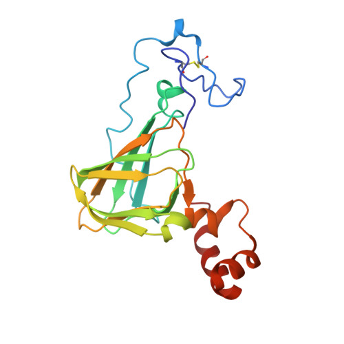
2ET7

Structural and spectroscopic insights into the mechanism of oxalate oxidase


Experimental Data Snapshot

  • Method: X-RAY DIFFRACTION
  • Resolution: 1.70 Å
  • R-Value Free: 0.211 
  • R-Value Work: 0.163 
  • R-Value Observed: 0.165 

wwPDB Validation   3D Report Full Report


This is version 1.5 of the entry. See complete history


Literature

Structural and spectroscopic studies shed light on the mechanism of oxalate oxidase

Opaleye, O.Rose, R.-S.Whittaker, M.M.Woo, E.-J.Whittaker, J.W.Pickersgill, R.W.

(2006) J Biol Chem 281: 6428-6433

  • DOI: https://doi.org/10.1074/jbc.M510256200
  • Primary Citation of Related Structures:  
    2ET1, 2ET7, 2ETE

  • PubMed Abstract: 

    Oxalate oxidase (EC 1.2.3.4) catalyzes the conversion of oxalate and dioxygen to hydrogen peroxide and carbon dioxide. In this study, glycolate was used as a structural analogue of oxalate to investigate substrate binding in the crystalline enzyme. The observed monodentate binding of glycolate to the active site manganese ion of oxalate oxidase is consistent with a mechanism involving C-C bond cleavage driven by superoxide anion attack on a monodentate coordinated substrate. In this mechanism, the metal serves two functions: to organize the substrates (oxalate and dioxygen) and to transiently reduce dioxygen. The observed structure further implies important roles for specific active site residues (two asparagines and one glutamine) in correctly orientating the substrates and reaction intermediates for catalysis. Combined spectroscopic, biochemical, and structural analyses of mutants confirms the importance of the asparagine residues in organizing a functional active site complex.


  • Organizational Affiliation

    School of Biological and Chemical Sciences, Queen Mary, University of London, Mile End Road, London E1 4NS, United Kingdom.


Macromolecules
Find similar proteins by:  (by identity cutoff)  |  3D Structure
Entity ID: 1
MoleculeChains Sequence LengthOrganismDetailsImage
Oxalate oxidase 1201Hordeum vulgareMutation(s): 1 
EC: 1.2.3.4
UniProt
Find proteins for P45850 (Hordeum vulgare)
Explore P45850 
Go to UniProtKB:  P45850
Entity Groups  
Sequence Clusters30% Identity50% Identity70% Identity90% Identity95% Identity100% Identity
UniProt GroupP45850
Sequence Annotations
Expand
  • Reference Sequence
Small Molecules
Ligands 1 Unique
IDChains Name / Formula / InChI Key2D Diagram3D Interactions
MN
Query on MN

Download Ideal Coordinates CCD File 
B [auth A]MANGANESE (II) ION
Mn
WAEMQWOKJMHJLA-UHFFFAOYSA-N
Experimental Data & Validation

Experimental Data

  • Method: X-RAY DIFFRACTION
  • Resolution: 1.70 Å
  • R-Value Free: 0.211 
  • R-Value Work: 0.163 
  • R-Value Observed: 0.165 
  • Space Group: H 3 2
Unit Cell:
Length ( Å )Angle ( ˚ )
a = 94.823α = 90
b = 94.823β = 90
c = 106.238γ = 120
Software Package:
Software NamePurpose
REFMACrefinement
MOSFLMdata reduction
CCP4data scaling

Structure Validation

View Full Validation Report


Entry History 

Deposition Data

Revision History  (Full details and data files)

  • Version 1.0: 2005-11-22
    Type: Initial release
  • Version 1.1: 2008-05-01
    Changes: Version format compliance
  • Version 1.2: 2011-07-13
    Changes: Derived calculations, Version format compliance
  • Version 1.3: 2017-10-18
    Changes: Refinement description
  • Version 1.4: 2021-10-20
    Changes: Database references, Derived calculations
  • Version 1.5: 2024-11-20
    Changes: Data collection, Structure summary