2EEG | pdb_00002eeg

Solution Structure of PDZ domain of PDZ and LIM domain protein


Experimental Data Snapshot

  • Method: SOLUTION NMR
  • Conformers Calculated: 100 
  • Conformers Submitted: 20 
  • Selection Criteria: structures with the least restraint violations, target function 

wwPDB Validation 3D Report Full Report

Validation slider image for 2EEG

This is version 1.3 of the entry. See complete history

Literature

Solution Structure of PDZ domain of PDZ and LIM domain protein

Niraula, T.N.Tomizawa, T.Koshiba, K.Inoue, M.Kigawa, T.Yokoyama, S.

To be published.

Macromolecule Content 

  • Total Structure Weight: 9.6 kDa 
  • Atom Count: 671 
  • Modeled Residue Count: 94 
  • Deposited Residue Count: 94 
  • Unique protein chains: 1

Macromolecules

Find similar proteins by:|  3D Structure
Entity ID: 1
MoleculeChains  Sequence LengthOrganismDetailsImage
PDZ and LIM domain protein 494Homo sapiensMutation(s): 0 
Gene Names: PDLIM4
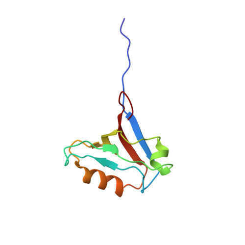
UniProt & NIH Common Fund Data Resources
Find proteins for P50479 (Homo sapiens)
Explore P50479 
Go to UniProtKB:  P50479
PHAROS:  P50479
GTEx:  ENSG00000131435 
Entity Groups
Sequence Clusters30% Identity50% Identity70% Identity90% Identity95% Identity100% Identity
UniProt GroupP50479
Sequence Annotations
Expand
Reference Sequence

Experimental Data & Validation

Experimental Data

  • Method: SOLUTION NMR
  • Conformers Calculated: 100 
  • Conformers Submitted: 20 
  • Selection Criteria: structures with the least restraint violations, target function 

Structure Validation

View Full Validation Report


Entry History 

Revision History  (Full details and data files)

  • Version 1.0: 2007-08-21
    Type: Initial release
  • Version 1.1: 2011-07-13
    Changes: Version format compliance
  • Version 1.2: 2022-03-09
    Changes: Data collection, Database references, Derived calculations
  • Version 1.3: 2024-05-29
    Changes: Data collection