2E5M | pdb_00002e5m

Crystal structure of isocitrate dehydrogenase from Sulfolobus tokodaii strain 7


Experimental Data Snapshot

  • Method: X-RAY DIFFRACTION
  • Resolution: 2.40 Å
  • R-Value Free: 
    0.279 (Depositor), 0.279 (DCC) 
  • R-Value Work: 
    0.217 (Depositor), 0.217 (DCC) 
  • R-Value Observed: 
    0.217 (Depositor) 

Starting Model: experimental
View more details

wwPDB Validation 3D Report Full Report

Validation slider image for 2E5M

Ligand Structure Quality Assessment 


This is version 1.4 of the entry. See complete history

Literature

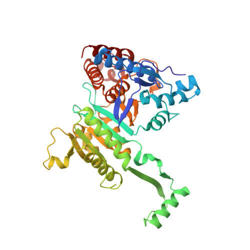
Crystal Structures of the Putative Isocitrate Dehydrogenase fromSulfolobus tokodaiiStrain 7 in the Apo and NADP+-Bound Forms.

Kondo, H.Murakami, M.

(2018) Archaea 2018: 7571984-7571984

  • DOI: https://doi.org/10.1155/2018/7571984
  • Primary Citation Related Structures: 
    2E0C, 2E5M

  • PubMed Abstract: 

    Isocitrate dehydrogenase is a catabolic enzyme that acts during the third step of the tricarboxylic acid cycle. The hypothetical protein ST2166 from the archaeon Sulfolobus tokodaii was isolated and crystallized. It shares high primary structure homology with prokaryotic NADP + -dependent IDHs, suggesting that these enzymes share a common enzymatic mechanism. The crystal structure of ST2166 was determined at 2.0 Å resolution in the apo form, and then the structure of the crystal soaked with NADP + was also determined at 2.4 Å resolution, which contained NADP + bound at the putative active site. Comparisons between the structures of apo and NADP + -bound forms and NADP-IDHs from other prokaryotes suggest that prokaryotic NADP-IDHs recognize their cofactors using conserved Lys335, Tyr336, and Arg386 in ST2166 at the opening cleft before the domain closure.


  • Organizational Affiliation
    • Department of Physics, Graduate School of Science, Nagoya University, 464-8602 Nagoya, Japan.

Macromolecule Content 

  • Total Structure Weight: 94.6 kDa 
  • Atom Count: 6,947 
  • Modeled Residue Count: 806 
  • Deposited Residue Count: 818 
  • Unique protein chains: 1

Macromolecules

Find similar proteins by:|  3D Structure
Entity ID: 1
MoleculeChains  Sequence LengthOrganismDetailsImage
409aa long hypothetical NADP-dependent isocitrate dehydrogenase
A, B
409Sulfurisphaera tokodaii str. 7Mutation(s): 0 
EC: 1.1.1.42
UniProt
Find proteins for Q96YK6 (Sulfurisphaera tokodaii (strain DSM 16993 / JCM 10545 / NBRC 100140 / 7))
Explore Q96YK6 
Go to UniProtKB:  Q96YK6
Entity Groups
Sequence Clusters30% Identity50% Identity70% Identity90% Identity95% Identity100% Identity
UniProt GroupQ96YK6
Sequence Annotations
Expand
Reference Sequence

Small Molecules

Experimental Data & Validation

Experimental Data

  • Method: X-RAY DIFFRACTION
  • Resolution: 2.40 Å
  • R-Value Free:  0.279 (Depositor), 0.279 (DCC) 
  • R-Value Work:  0.217 (Depositor), 0.217 (DCC) 
  • R-Value Observed: 0.217 (Depositor) 
Space Group: P 1 21 1
Unit Cell:
Length ( Å )Angle ( ˚ )
a = 74.78α = 90
b = 88.11β = 91.36
c = 75.67γ = 90
Software Package:
Software NamePurpose
ADSCdata collection
CNSrefinement
MOSFLMdata reduction
SCALAdata scaling
CNSphasing

Structure Validation

View Full Validation Report


Ligand Structure Quality Assessment 


Entry History 

Deposition Data

Revision History  (Full details and data files)

  • Version 1.0: 2008-01-15
    Type: Initial release
  • Version 1.1: 2011-07-13
    Changes: Derived calculations, Version format compliance
  • Version 1.2: 2019-01-23
    Changes: Data collection, Database references
  • Version 1.3: 2020-01-01
    Changes: Database references
  • Version 1.4: 2023-10-25
    Changes: Data collection, Database references, Derived calculations, Refinement description