2E24 | pdb_00002e24

crystal structure of a mutant (R612A) of xanthan lyase


Experimental Data Snapshot

  • Method: X-RAY DIFFRACTION
  • Resolution: 2.15 Å
  • R-Value Free: 
    0.217 (Depositor), 0.215 (DCC) 
  • R-Value Work: 
    0.175 (Depositor), 0.175 (DCC) 
  • R-Value Observed: 
    0.175 (Depositor) 

Starting Model: experimental
View more details

wwPDB Validation 3D Report Full Report

Validation slider image for 2E24

This is version 1.4 of the entry. See complete history

Literature

A Structural Factor Responsible for Substrate Recognition by Bacillus sp. GL1 Xanthan Lyase that Acts Specifically on Pyruvated Side Chains of Xanthan

Maruyama, Y.Mikami, B.Hashimoto, W.Murata, K.

(2007) Biochemistry 46: 781-791

  • DOI: https://doi.org/10.1021/bi0619775
  • Primary Citation Related Structures: 
    2E22, 2E24

  • PubMed Abstract: 

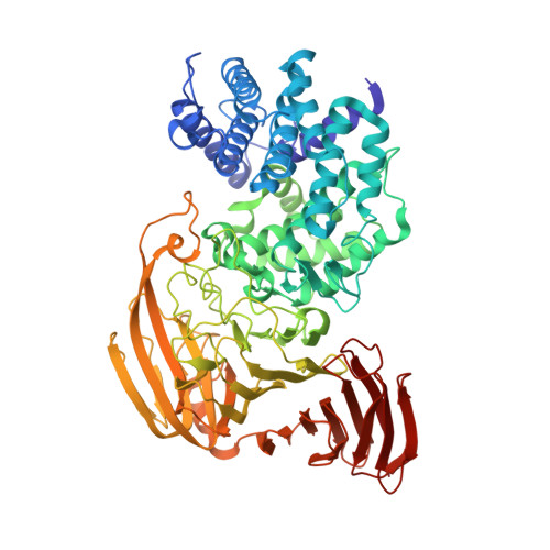
    Xanthan is a bacterial heteropolysaccharide composed of pentasaccharide repeating units, i.e., a cellobiose as a backbone and a trisaccharide consisting of two mannoses and one glucuronic acid as a side chain. Nonreducing terminal mannose residues of xanthan side chains are partially pyruvated. Bacillus sp. GL1 xanthan lyase, a member of polysaccharide lyase family 8, acts specifically on pyruvated side chains of xanthan and yields pyruvated mannose through a beta-elimination reaction by using a single Tyr255 residue as base and acid catalysts. Here we show structural factors for substrate recognition by xanthan lyase through X-ray crystallographic and mutational analyses. The enzyme accommodates mannose and pyruvated mannose at the -1 subsite, although both inhibitor and dissociation constants of the two monosaccharides indicated that the affinity of pyruvated mannose for xanthan lyase is much higher than that of mannose. The high affinity of pyruvated mannose is probably due to the formation of additional hydrogen bonds between the carboxyl group of pyruvated mannose and amino acid residues of Tyr315 and Arg612. Site-directed mutagenesis of the two residues demonstrated that Arg612 is a key residue in recognizing pyruvated mannose. Arg612 is located in the protruding loop covering the substrate, suggesting that the loop functions as a lid that is responsible for the proper accommodation of the substrate at the active site.


  • Organizational Affiliation
    • Laboratory of Basic and Applied Molecular Biotechnology, Division of Food Science and Biotechnology, Graduate School of Agriculture, Kyoto University, Uji, Kyoto 611-0011, Japan.

Macromolecule Content 

  • Total Structure Weight: 80.78 kDa 
  • Atom Count: 6,255 
  • Modeled Residue Count: 752 
  • Deposited Residue Count: 752 
  • Unique protein chains: 1

Macromolecules

Find similar proteins by:|  3D Structure
Entity ID: 1
MoleculeChains  Sequence LengthOrganismDetailsImage
Xanthan lyase752Bacillus sp. GL1Mutation(s): 1 
EC: 4.2.2.12
UniProt
Find proteins for Q9AQS0 (Bacillus sp. (strain GL1))
Explore Q9AQS0 
Go to UniProtKB:  Q9AQS0
Entity Groups
Sequence Clusters30% Identity50% Identity70% Identity90% Identity95% Identity100% Identity
UniProt GroupQ9AQS0
Sequence Annotations
Expand
Reference Sequence

Small Molecules

Ligands 1 Unique
IDChains Name / Formula / InChI Key2D Diagram3D Interactions
PEG

Query on PEG


Download:Ideal Coordinates CCD File
B [auth A]DI(HYDROXYETHYL)ETHER
C4 H10 O3
MTHSVFCYNBDYFN-UHFFFAOYSA-N

Experimental Data & Validation

Experimental Data

  • Method: X-RAY DIFFRACTION
  • Resolution: 2.15 Å
  • R-Value Free:  0.217 (Depositor), 0.215 (DCC) 
  • R-Value Work:  0.175 (Depositor), 0.175 (DCC) 
  • R-Value Observed: 0.175 (Depositor) 
Space Group: P 1 21 1
Unit Cell:
Length ( Å )Angle ( ˚ )
a = 53.807α = 90
b = 90.23β = 97.56
c = 78.098γ = 90
Software Package:
Software NamePurpose
CNSrefinement
HKL-2000data reduction
HKL-2000data scaling
CNSphasing

Structure Validation

View Full Validation Report


Entry History 

Deposition Data

Revision History  (Full details and data files)

  • Version 1.0: 2007-01-30
    Type: Initial release
  • Version 1.1: 2008-04-30
    Changes: Version format compliance
  • Version 1.2: 2011-07-13
    Changes: Source and taxonomy, Version format compliance
  • Version 1.3: 2021-11-10
    Changes: Database references, Derived calculations
  • Version 1.4: 2023-10-25
    Changes: Data collection, Refinement description