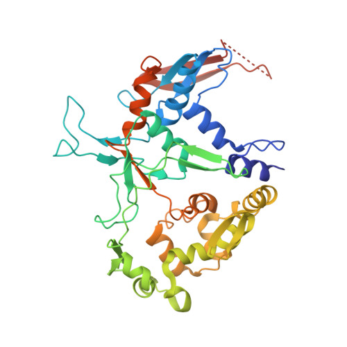
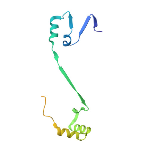
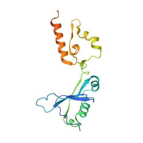
2E1M

Crystal Structure of L-Glutamate Oxidase from Streptomyces sp. X-119-6


Experimental Data Snapshot

  • Method: X-RAY DIFFRACTION
  • Resolution: 2.80 Å
  • R-Value Free: 0.308 
  • R-Value Work: 0.248 
  • R-Value Observed: 0.248 

Starting Model: experimental
View more details

wwPDB Validation   3D Report Full Report


Ligand Structure Quality Assessment 


This is version 1.2 of the entry. See complete history


Literature

Structural characterization of l-glutamate oxidase from Streptomyces sp. X-119-6

Arima, J.Sasaki, C.Sakaguchi, C.Mizuno, H.Tamura, T.Kashima, A.Kusakabe, H.Sugio, S.Inagaki, K.

(2009) FEBS J 276: 4318-4327

  • DOI: https://doi.org/10.1111/j.1742-4658.2009.07103.x
  • Primary Citation of Related Structures:  
    2E1M

  • PubMed Abstract: 

    L-Glutamate oxidase (LGOX) from Streptomyces sp. X-119-6, which catalyzes the oxidative deamination of L-glutamate, has attracted increasing attention as a component of amperometric L-glutamate sensors used in the food industry and clinical biochemistry. The precursor of LGOX, which has a homodimeric structure, is less active than the mature enzyme with an alpha(2)beta(2)V(2) structure; enzymatic proteolysis of the precursor forms the stable mature enzyme. We solved the crystal structure of mature LGOX using molecular replacement with a structurally homologous model of L-amino acid oxidase (LAAO) from snake venom: LGOX has a deeply buried active site and two entrances from the surface of the protein into the active site. Comparison of the LGOX structure with that of LAAO revealed that LGOX has three regions that are absent from the LAAO structure, one of which is involved in the formation of the entrance. Furthermore, the arrangement of the residues composing the active site differs between LGOX and LAAO, and the active site of LGOX is narrower than that of LAAO. Results of the comparative analyses described herein raise the possibility that such a unique structure of LGOX is associated with its substrate specificity.


  • Organizational Affiliation

    Department of Biofunctional Chemistry, Graduate School of Natural Science and Technology, Okayama University, Japan.


Macromolecules
Find similar proteins by:  (by identity cutoff)  |  3D Structure
Entity ID: 1
MoleculeChains Sequence LengthOrganismDetailsImage
L-glutamate oxidase376Streptomyces sp. X-119-6Mutation(s): 0 
EC: 1.4.3.11
UniProt
Find proteins for Q8L3C7 (Streptomyces sp)
Explore Q8L3C7 
Go to UniProtKB:  Q8L3C7
Entity Groups  
Sequence Clusters30% Identity50% Identity70% Identity90% Identity95% Identity100% Identity
UniProt GroupQ8L3C7
Sequence Annotations
Expand
  • Reference Sequence
Find similar proteins by:  (by identity cutoff)  |  3D Structure
Entity ID: 2
MoleculeChains Sequence LengthOrganismDetailsImage
L-glutamate oxidase130Streptomyces sp. X-119-6Mutation(s): 0 
EC: 1.4.3.11
UniProt
Find proteins for Q8L3C7 (Streptomyces sp)
Explore Q8L3C7 
Go to UniProtKB:  Q8L3C7
Entity Groups  
Sequence Clusters30% Identity50% Identity70% Identity90% Identity95% Identity100% Identity
UniProt GroupQ8L3C7
Sequence Annotations
Expand
  • Reference Sequence
Find similar proteins by:  (by identity cutoff)  |  3D Structure
Entity ID: 3
MoleculeChains Sequence LengthOrganismDetailsImage
L-glutamate oxidase181Streptomyces sp. X-119-6Mutation(s): 0 
EC: 1.4.3.11
UniProt
Find proteins for Q8L3C7 (Streptomyces sp)
Explore Q8L3C7 
Go to UniProtKB:  Q8L3C7
Entity Groups  
Sequence Clusters30% Identity50% Identity70% Identity90% Identity95% Identity100% Identity
UniProt GroupQ8L3C7
Sequence Annotations
Expand
  • Reference Sequence
Experimental Data & Validation

Experimental Data

  • Method: X-RAY DIFFRACTION
  • Resolution: 2.80 Å
  • R-Value Free: 0.308 
  • R-Value Work: 0.248 
  • R-Value Observed: 0.248 
  • Space Group: P 61 2 2
Unit Cell:
Length ( Å )Angle ( ˚ )
a = 123.875α = 90
b = 123.875β = 90
c = 168.762γ = 120
Software Package:
Software NamePurpose
HKL-2000data collection
AMoREphasing
CNSrefinement
HKL-2000data reduction
HKL-2000data scaling

Structure Validation

View Full Validation Report


Ligand Structure Quality Assessment 


Entry History 

Deposition Data

Revision History  (Full details and data files)

  • Version 1.0: 2007-11-13
    Type: Initial release
  • Version 1.1: 2011-07-13
    Changes: Derived calculations, Source and taxonomy, Version format compliance
  • Version 1.2: 2023-10-25
    Changes: Data collection, Database references, Derived calculations, Refinement description