A Third Mode of DNA Binding: Phosphate Clamps by a Polynuclear Platinum Complex
Komeda, S., Moulaei, T., Woods, K.K., Chikuma, M., Farrell, N.P., Williams, L.D.(2006) J Am Chem Soc 128: 16092-16103
- PubMed: 17165762 Search on PubMed
- DOI: https://doi.org/10.1021/ja062851y
- Primary Citation Related Structures:
2DYW - PubMed Abstract:
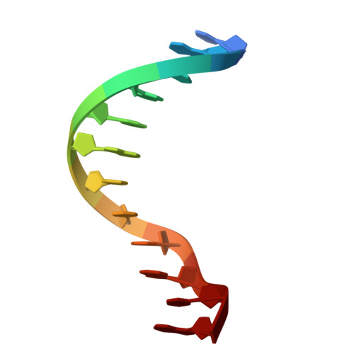
We describe a 1.2 A X-ray structure of a double-stranded B-DNA dodecamer (the Dickerson Dodecamer, DDD, [d(CGCGAATTCGCG)]2) associated with a cytotoxic platinum(II) complex, [{trans-Pt(NH3)2(NH2(CH2)6(NH3+)}2-mu-{trans-Pt(NH3)2(NH2(CH2)6NH2)2}] (TriplatinNC). TriplatinNC is a multifunctional DNA ligand, with three cationic Pt(II) centers, and directional hydrogen bonding functionalities, linked by flexible hydrophobic segments, but without the potential for covalent interaction. TriplatinNC does not intercalate nor does it bind in either groove. Instead, it binds to phosphate oxygen atoms and thus associates with the backbone. The three square-planar tetra-am(m)ine Pt(II) coordination units form bidentate N...O...N complexes with OP atoms, in a motif we call the Phosphate Clamp. The geometry is conserved among the 8 observed phosphate clamps in this structure. The interaction appears to prefer O2P over O1P atoms (frequency of interaction is O2P > O1P, base and sugar oxygens > N). The high repetition and geometric regularity of the motif suggests that this type of Pt(II) center can be developed as a modular nucleic acid binding device with general utility. TriplatinNC extends along the phosphate backbone, in a mode of binding we call "Backbone Tracking" and spans the minor groove in a mode of binding we call "Groove Spanning". Electrostatic forces appear to induce modest DNA bending into the major groove. This bending may be related to the direct coordination of a sodium cation by a DNA base, with unprecedented inner-shell (direct) coordination of penta-hydrated sodium at the O6 atom of a guanine.
- School of Chemistry and Biochemistry, Georgia Institute of Technology, Atlanta, Georgia 30332-0400, USA.
Organizational Affiliation: