2DY5 | pdb_00002dy5

Crystal structure of rat heme oxygenase-1 in complex with heme and 2-[2-(4-chlorophenyl)ethyl]-2-[(1H-imidazol-1-yl)methyl]-1,3-dioxolane


Experimental Data Snapshot

  • Method: X-RAY DIFFRACTION
  • Resolution: 2.70 Å
  • R-Value Free: 
    0.248 (Depositor), 0.234 (DCC) 
  • R-Value Work: 
    0.194 (Depositor), 0.188 (DCC) 
  • R-Value Observed: 
    0.197 (Depositor) 

Starting Model: experimental
View more details

wwPDB Validation 3D Report Full Report

Validation slider image for 2DY5

Ligand Structure Quality Assessment 


This is version 1.3 of the entry. See complete history

Literature

X-ray crystallographic and biochemical characterization of the inhibitory action of an imidazole-dioxolane compound on heme oxygenase

Sugishima, M.Higashimoto, Y.Oishi, T.Takahashi, H.Sakamoto, H.Noguchi, M.Fukuyama, K.

(2007) Biochemistry 46: 1860-1867

  • DOI: https://doi.org/10.1021/bi062264p
  • Primary Citation Related Structures: 
    2DY5

  • PubMed Abstract: 

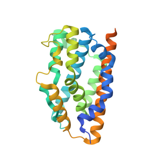
    Heme oxygenase (HO) catalyzes the regiospecific cleavage of the porphyrin ring of heme using reducing equivalents and O2 to produce biliverdin, iron, and CO. Because CO has a cytoprotective effect through the p38-MAPK pathway, HO is a potential therapeutic target in cancer. In fact, inhibition of the HO isoform HO-1 reduces Kaposi sarcoma tumor growth. Imidazole-dioxolane compounds have recently attracted attention because they have been reported to specifically inhibit HO-1, but not HO-2, unlike Cr-containing protoporphyrin IX, a classical inhibitor of HO, that inhibits not only both HO isoforms but also other hemoproteins. The inhibitory mechanism of imidazole-dioxolane compounds, however, has not yet been characterized. Here, we determine the crystal structure of the ternary complex of rat HO-1, heme, and an imidazole-dioxolane compound, 2-[2-(4-chlorophenyl)ethyl]-2-[(1H-imidazol-1-yl)methyl]-1,3-dioxolane. This compound bound on the distal side of the heme iron, where the imidazole and 4-chlorophenyl groups were bound to the heme iron and the hydrophobic cavity in HO, respectively. Binding of the bulky inhibitor in the narrow distal pocket shifted the distal helix to open the distal site and moved both the heme and the proximal helix. Furthermore, the biochemical characterization revealed that the catalytic reactions of both HO-1 and HO-2 were completely stopped after the formation of verdoheme in the presence of the imidazole-dioxolane compound. This result should be mainly due to the lower reactivity of the inhibitor-bound verdoheme with O2 compared to the reactivity of the inhibitor-bound heme with O2.


  • Organizational Affiliation
    • Department of Medical Biochemistry, Kurume University School of Medicine, Kurume, Fukuoka 830-0011, Japan.

Macromolecule Content 

  • Total Structure Weight: 31.56 kDa 
  • Atom Count: 1,816 
  • Modeled Residue Count: 214 
  • Deposited Residue Count: 267 
  • Unique protein chains: 1

Macromolecules

Find similar proteins by:|  3D Structure
Entity ID: 1
MoleculeChains  Sequence LengthOrganismDetailsImage
Heme oxygenase 1267Rattus norvegicusMutation(s): 0 
EC: 1.14.99.3 (PDB Primary Data), 1.14.14.18 (UniProt)
UniProt
Find proteins for P06762 (Rattus norvegicus)
Explore P06762 
Go to UniProtKB:  P06762
Entity Groups
Sequence Clusters30% Identity50% Identity70% Identity90% Identity95% Identity100% Identity
UniProt GroupP06762
Sequence Annotations
Expand
Reference Sequence

Small Molecules

Ligands 3 Unique
IDChains Name / Formula / InChI Key2D Diagram3D Interactions
HEM

Query on HEM


Download:Ideal Coordinates CCD File
C [auth A]PROTOPORPHYRIN IX CONTAINING FE
C34 H32 Fe N4 O4
KABFMIBPWCXCRK-RGGAHWMASA-L
224

Query on 224


Download:Ideal Coordinates CCD File
D [auth A]1-({2-[2-(4-CHLOROPHENYL)ETHYL]-1,3-DIOXOLAN-2-YL}METHYL)-1H-IMIDAZOLE
C15 H17 Cl N2 O2
NPIOYRIZNLPLDH-UHFFFAOYSA-N
CL

Query on CL


Download:Ideal Coordinates CCD File
B [auth A]CHLORIDE ION
Cl
VEXZGXHMUGYJMC-UHFFFAOYSA-M
Binding Affinity Annotations 
IDSourceBinding Affinity
224 BindingDB:  2DY5 IC50: min: 2800, max: 4300 (nM) from 3 assay(s)

Experimental Data & Validation

Experimental Data

  • Method: X-RAY DIFFRACTION
  • Resolution: 2.70 Å
  • R-Value Free:  0.248 (Depositor), 0.234 (DCC) 
  • R-Value Work:  0.194 (Depositor), 0.188 (DCC) 
  • R-Value Observed: 0.197 (Depositor) 
Space Group: P 32 2 1
Unit Cell:
Length ( Å )Angle ( ˚ )
a = 66.78α = 90
b = 66.78β = 90
c = 121.39γ = 120
Software Package:
Software NamePurpose
HKL-2000data collection
MOLREPphasing
CNSrefinement
HKL-2000data reduction
HKL-2000data scaling

Structure Validation

View Full Validation Report


Ligand Structure Quality Assessment 


Entry History 

Deposition Data

Revision History  (Full details and data files)

  • Version 1.0: 2007-05-22
    Type: Initial release
  • Version 1.1: 2008-04-30
    Changes: Version format compliance
  • Version 1.2: 2011-07-13
    Changes: Version format compliance
  • Version 1.3: 2023-10-25
    Changes: Data collection, Database references, Derived calculations, Refinement description