2DUR | pdb_00002dur

Crystal structure of VIP36 exoplasmic/lumenal domain, Ca2+/Man2-bound form


Experimental Data Snapshot

  • Method: X-RAY DIFFRACTION
  • Resolution: 1.65 Å
  • R-Value Free: 
    0.228 (Depositor), 0.231 (DCC) 
  • R-Value Work: 
    0.199 (Depositor), 0.203 (DCC) 
  • R-Value Observed: 
    0.200 (Depositor) 

Starting Model: experimental
View more details

wwPDB Validation 3D Report Full Report

Validation slider image for 2DUR

This is version 2.2 of the entry. See complete history

Literature

Structural basis for recognition of high mannose type glycoproteins by mammalian transport lectin VIP36

Satoh, T.Cowieson, N.P.Hakamata, W.Ideo, H.Fukushima, K.Kurihara, M.Kato, R.Yamashita, K.Wakatsuki, S.

(2007) J Biological Chem 282: 28246-28255

  • DOI: https://doi.org/10.1074/jbc.M703064200
  • Primary Citation Related Structures: 
    2DUO, 2DUP, 2DUQ, 2DUR, 2E6V

  • PubMed Abstract: 

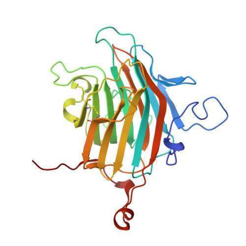
    VIP36 functions as a transport lectin for trafficking certain high mannose type glycoproteins in the secretory pathway. Here we report the crystal structure of VIP36 exoplasmic/luminal domain comprising a carbohydrate recognition domain and a stalk domain. The structures of VIP36 in complex with Ca(2+) and mannosyl ligands are also described. The carbohydrate recognition domain is composed of a 17-stranded antiparallel beta-sandwich and binds one Ca(2+) adjoining the carbohydrate-binding site. The structure reveals that a coordinated Ca(2+) ion orients the side chains of Asp(131), Asn(166), and His(190) for carbohydrate binding. This result explains the Ca(2+)-dependent carbohydrate binding of this protein. The Man-alpha-1,2-Man-alpha-1,2-Man, which corresponds to the D1 arm of high mannose type glycan, is recognized by eight residues through extensive hydrogen bonds. The complex structures reveal the structural basis for high mannose type glycoprotein recognition by VIP36 in a Ca(2+)-dependent and D1 arm-specific manner.


  • Organizational Affiliation
    • Structural Biology Research Center, Photon Factory, Institute of Materials Structure Science, High Energy Accelerator Research Organization (KEK), Tsukuba, Ibaraki 305-0801, Japan.

Macromolecule Content 

  • Total Structure Weight: 59.1 kDa 
  • Atom Count: 4,557 
  • Modeled Residue Count: 501 
  • Deposited Residue Count: 506 
  • Unique protein chains: 1

Macromolecules

Find similar proteins by:|  3D Structure
Entity ID: 1
MoleculeChains  Sequence LengthOrganismDetailsImage
Vesicular integral-membrane protein VIP36
A, B
253Canis lupus familiarisMutation(s): 0 
Gene Names: LMAN2
UniProt
Find proteins for P49256 (Canis lupus familiaris)
Explore P49256 
Go to UniProtKB:  P49256
Entity Groups
Sequence Clusters30% Identity50% Identity70% Identity90% Identity95% Identity100% Identity
UniProt GroupP49256
Sequence Annotations
Expand
Reference Sequence

Oligosaccharides

Help  
Entity ID: 2
MoleculeChains Length2D Diagram GlycosylationD Interactions
alpha-D-mannopyranose-(1-2)-alpha-D-mannopyranose
C, D
2N/A
Glycosylation Resources
GlyTouCan: G53402KW
GlyCosmos: G53402KW
GlyGen: G53402KW

Small Molecules

Ligands 3 Unique
IDChains Name / Formula / InChI Key2D Diagram3D Interactions
GOL

Query on GOL


Download:Ideal Coordinates CCD File
K [auth A],
L [auth A]
GLYCEROL
C3 H8 O3
PEDCQBHIVMGVHV-UHFFFAOYSA-N
CA

Query on CA


Download:Ideal Coordinates CCD File
E [auth A],
M [auth B]
CALCIUM ION
Ca
BHPQYMZQTOCNFJ-UHFFFAOYSA-N
CL

Query on CL


Download:Ideal Coordinates CCD File
F [auth A]
G [auth A]
H [auth A]
I [auth A]
J [auth A]
F [auth A],
G [auth A],
H [auth A],
I [auth A],
J [auth A],
N [auth B],
O [auth B],
P [auth B]
CHLORIDE ION
Cl
VEXZGXHMUGYJMC-UHFFFAOYSA-M

Biologically Interesting Molecules (External Reference) 

1 Unique

Experimental Data & Validation

Experimental Data

  • Method: X-RAY DIFFRACTION
  • Resolution: 1.65 Å
  • R-Value Free:  0.228 (Depositor), 0.231 (DCC) 
  • R-Value Work:  0.199 (Depositor), 0.203 (DCC) 
  • R-Value Observed: 0.200 (Depositor) 
Space Group: C 1 2 1
Unit Cell:
Length ( Å )Angle ( ˚ )
a = 171.2α = 90
b = 45.5β = 132.7
c = 117.4γ = 90
Software Package:
Software NamePurpose
REFMACrefinement
DENZOdata reduction
SCALEPACKdata scaling
MOLREPphasing

Structure Validation

View Full Validation Report


Entry History 

Deposition Data

Revision History  (Full details and data files)

  • Version 1.0: 2007-07-24
    Type: Initial release
  • Version 1.1: 2007-10-22
    Changes: Version format compliance
  • Version 1.2: 2011-07-13
    Changes: Non-polymer description, Version format compliance
  • Version 2.0: 2020-07-29
    Type: Remediation
    Reason: Carbohydrate remediation
    Changes: Atomic model, Data collection, Database references, Derived calculations, Structure summary
  • Version 2.1: 2023-10-25
    Changes: Data collection, Database references, Refinement description, Structure summary
  • Version 2.2: 2024-11-20
    Changes: Structure summary