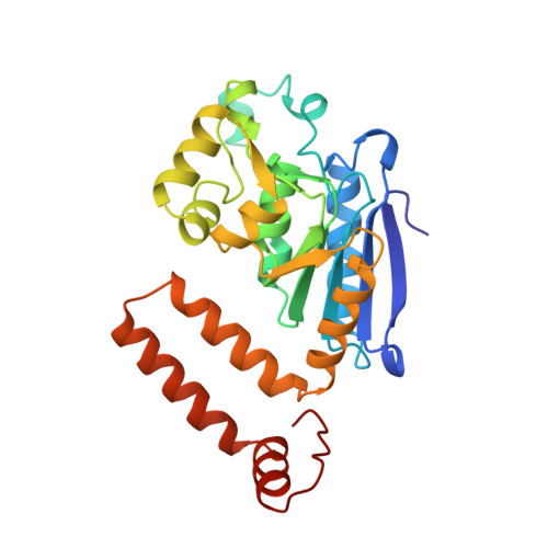
The crystal structure of enoyl-CoA hydratase complexed with octanoyl-CoA reveals the structural adaptations required for binding of a long chain fatty acid-CoA molecule.
Engel, C.K., Kiema, T.R., Hiltunen, J.K., Wierenga, R.K.(1998) J Mol Biology 275: 847-859
- PubMed: 9480773 Search on PubMed
- DOI: https://doi.org/10.1006/jmbi.1997.1491
- Primary Citation Related Structures:
2DUB - PubMed Abstract:
The structure of the hexameric rat mitochondrial enoyl-Coenzyme A (CoA) hydratase, co-crystallised with the inhibitor octanoyl-CoA, has been refined at a resolution of 2.4 A. Enoyl-CoA hydratase catalyses the hydration of 2,3-unsaturated enoyl-CoA thioesters. In the crystal structure only four of the six active sites of the hexamer in the asymmetric unit are occupied with a ligand molecule, showing an unliganded and a liganded active site within the same crystal form. While the protein assembly and fold is identical to the previously solved acetoacetyl-CoA complex, differences are observed close to the fatty acid binding pocket due to the different nature of the ligands. The fatty acid tail of octanoyl-CoA is bound in an extended conformation. This is possible because a high B-factor loop, which separates in the acetoacetyl-CoA complex the binding pocket of the acetoacetyl-CoA fatty acid tail from the intertrimer space, has moved aside to allow binding of the longer octanoyl-CoA moiety. The movement of this loop opens a tunnel which traverses the complete subunit from the solvent space to the intertrimer space. The conformation of the catalytic residues is identical, in both structures as well as in the liganded and the unliganded active sites. In the unliganded active sites a water molecules is bound between the two catalytic glutamate, residues Glu144 and Glu164. After superposition of a liganded active site on an unliganded active site this water molecule is close to the carbon centre that becomes hydroxylated in the hydratase reaction. These findings support the idea that the active site is rigid and that the catalytic residues and the water molecule, as seen in the unliganded active site, are pre-positioned for very efficient catalysis.
- European Molecular Biology Laboratory, Heidelberg, D69012, Germany.
Organizational Affiliation: