2DRO

Crystal structure of reducing-end-xylose releasing exo-oligoxylanase D263C mutant


Experimental Data Snapshot

  • Method: X-RAY DIFFRACTION
  • Resolution: 1.70 Å
  • R-Value Free: 0.187 
  • R-Value Work: 0.161 
  • R-Value Observed: 0.162 

wwPDB Validation   3D Report Full Report


This is version 1.4 of the entry. See complete history


Literature

Structural explanation for the acquisition of glycosynthase activity

Hidaka, M.Fushinobu, S.Honda, Y.Wakagi, T.Shoun, H.Kitaoka, M.

(2009) J Biochem 

  • DOI: https://doi.org/10.1093/jb/mvp159
  • Primary Citation of Related Structures:  
    2DRO, 2DRQ, 2DRR, 2DRS, 3A3V

  • PubMed Abstract: 

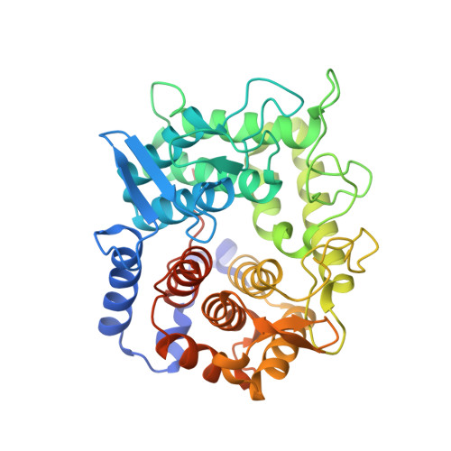
    Glycosynthases are engineered glycoside hydrolases (GHs) that catalyse the synthesis of glycoside from glycosyl-fluoride donors and suitable acceptors. We have determined five crystal structures of the glycosynthase mutants reducing-end xylose-releasing exo-oligoxylanase, an inverting GH, that exhibit various levels of glycosynthetic activities. At the active site of the Y198F mutant, the most efficient glycosynthase, a water molecule is observed at the same position as nucleophilic water (NW) in the parent enzyme, and the loss of the fixation of the direction of the lone pair of water molecules in the mutant drastically decreases hydrolytic activity. Water molecules were also observed at each active site of the general base mutant, but they were shifted 1.0-3.0 A from the NW in the wild type. Their positions exhibited a strong correlation with the strength of glycosynthase activity. Here, we propose that a structural prerequisite for the sufficient glycosynthase reaction is the presence of a water molecule at the NW position, and mutation at the NW holder provides a general strategy for inverting GHs. The idea on the position of a water molecule may also be applicable to the design of efficient glycosynthases from retaining GHs.


  • Organizational Affiliation

    Department of Biotechnology, The University of Tokyo, Tokyo, Japan.


Macromolecules
Find similar proteins by:  (by identity cutoff)  |  3D Structure
Entity ID: 1
MoleculeChains Sequence LengthOrganismDetailsImage
Xylanase Y396Halalkalibacterium halodurans C-125Mutation(s): 2 
Gene Names: BH2105
EC: 3.2.1.156
UniProt
Find proteins for Q9KB30 (Halalkalibacterium halodurans (strain ATCC BAA-125 / DSM 18197 / FERM 7344 / JCM 9153 / C-125))
Explore Q9KB30 
Go to UniProtKB:  Q9KB30
Entity Groups  
Sequence Clusters30% Identity50% Identity70% Identity90% Identity95% Identity100% Identity
UniProt GroupQ9KB30
Sequence Annotations
Expand
  • Reference Sequence
Experimental Data & Validation

Experimental Data

  • Method: X-RAY DIFFRACTION
  • Resolution: 1.70 Å
  • R-Value Free: 0.187 
  • R-Value Work: 0.161 
  • R-Value Observed: 0.162 
  • Space Group: P 21 21 21
Unit Cell:
Length ( Å )Angle ( ˚ )
a = 53.242α = 90
b = 86.369β = 90
c = 87.653γ = 90
Software Package:
Software NamePurpose
REFMACrefinement
ADSCdata collection
HKL-2000data scaling

Structure Validation

View Full Validation Report


Entry History 

Deposition Data

Revision History  (Full details and data files)

  • Version 1.0: 2006-06-27
    Type: Initial release
  • Version 1.1: 2008-04-30
    Changes: Version format compliance
  • Version 1.2: 2011-07-13
    Changes: Source and taxonomy, Version format compliance
  • Version 1.3: 2021-11-10
    Changes: Database references, Derived calculations
  • Version 1.4: 2023-10-25
    Changes: Data collection, Refinement description