2DQJ | pdb_00002dqj

Crystal structure of hyhel-10 FV (wild-type) complexed with hen egg lysozyme at 1.8A resolution


Experimental Data Snapshot

  • Method: X-RAY DIFFRACTION
  • Resolution: 1.80 Å
  • R-Value Free: 
    0.219 (Depositor), 0.219 (DCC) 
  • R-Value Work: 
    0.194 (Depositor), 0.193 (DCC) 
  • R-Value Observed: 
    0.194 (Depositor) 

Starting Model: experimental
View more details

wwPDB Validation 3D Report Full Report

Validation slider image for 2DQJ

This is version 1.4 of the entry. See complete history

Literature

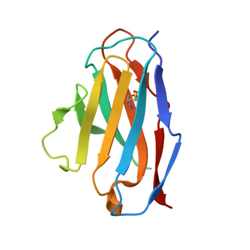
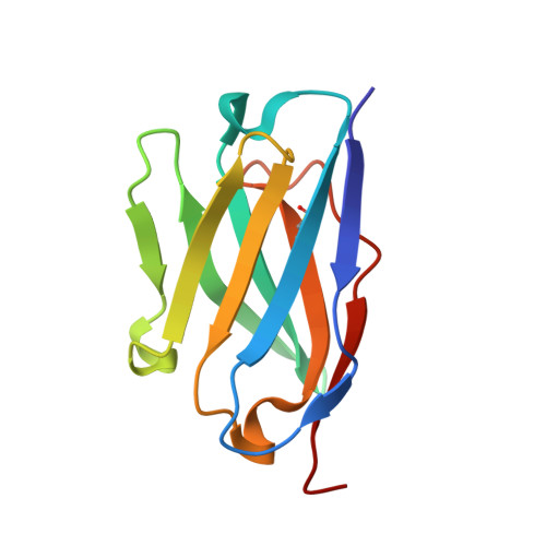
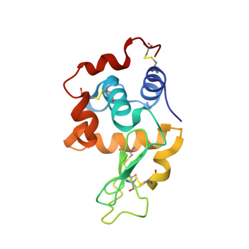
Structural consequences of mutations in interfacial Tyr residues of a protein antigen-antibody complex. The case of HyHEL-10-HEL

Shiroishi, M.Tsumoto, K.Tanaka, Y.Yokota, A.Nakanishi, T.Kondo, H.Kumagai, I.

(2007) J Biological Chem 282: 6783-6791

  • DOI: https://doi.org/10.1074/jbc.M605197200
  • Primary Citation Related Structures: 
    2DQC, 2DQD, 2DQE, 2DQF, 2DQG, 2DQH, 2DQI, 2DQJ

  • PubMed Abstract: 

    Tyrosine is an important amino acid in protein-protein interaction hot spots. In particular, many Tyr residues are located in the antigen-binding sites of antibodies and endow high affinity and high specificity to these antibodies. To investigate the role of interfacial Tyr residues in protein-protein interactions, we performed crystallographic studies and thermodynamic analyses of the interaction between hen egg lysozyme (HEL) and the anti-HEL antibody HyHEL-10 Fv fragment. HyHEL-10 has six Tyr residues in its antigen-binding site, which were systematically mutated to Phe and Ala using site-directed mutagenesis. The crystal structures revealed several critical roles for these Tyr residues in the interaction between HEL and HyHEL-10 as follows: 1) the aromatic ring of Tyr-50 in the light chain (LTyr-50) was important for the correct ternary structure of variable regions of the immunoglobulin light chain and heavy chain and of HEL; 2) deletion of the hydroxyl group of Tyr-50 in the heavy chain (HTyr-50) resulted in structural changes in the antigen-antibody interface; and 3) the side chains of HTyr-33 and HTyr-53 may help induce fitting of the antibody to the antigen. Hot spot Tyr residues may contribute to the high affinity and high specificity of the antigen-antibody interaction through a diverse set of structural and thermodynamic interactions.


  • Organizational Affiliation
    • Department of Biomolecular Engineering, Graduate School of Engineering, Tohoku University, Aoba-yama 6-6-11, Sendai 980-8579, Japan.

Macromolecule Content 

  • Total Structure Weight: 38.75 kDa 
  • Atom Count: 3,051 
  • Modeled Residue Count: 350 
  • Deposited Residue Count: 350 
  • Unique protein chains: 3

Macromolecules

Find similar proteins by:|  3D Structure
Entity ID: 1
MoleculeChains  Sequence LengthOrganismDetailsImage
lysozyme binding Ig kappa chain V23-J2 regionA [auth L]107Mus musculusMutation(s): 0 
Find similar proteins by:|  3D Structure
Entity ID: 2
MoleculeChains  Sequence LengthOrganismDetailsImage
Ig VH,anti-lysozymeB [auth H]114Mus musculusMutation(s): 0 
UniProt
Find proteins for P01823 (Mus musculus)
Explore P01823 
Go to UniProtKB:  P01823
Entity Groups
Sequence Clusters30% Identity50% Identity70% Identity90% Identity95% Identity100% Identity
UniProt GroupP01823
Sequence Annotations
Expand
Reference Sequence
Find similar proteins by:|  3D Structure
Entity ID: 3
MoleculeChains  Sequence LengthOrganismDetailsImage
Lysozyme CC [auth Y]129Gallus gallusMutation(s): 0 
EC: 3.2.1.17
UniProt
Find proteins for P00698 (Gallus gallus)
Explore P00698 
Go to UniProtKB:  P00698
Entity Groups
Sequence Clusters30% Identity50% Identity70% Identity90% Identity95% Identity100% Identity
UniProt GroupP00698
Sequence Annotations
Expand
Reference Sequence

Experimental Data & Validation

Experimental Data

  • Method: X-RAY DIFFRACTION
  • Resolution: 1.80 Å
  • R-Value Free:  0.219 (Depositor), 0.219 (DCC) 
  • R-Value Work:  0.194 (Depositor), 0.193 (DCC) 
  • R-Value Observed: 0.194 (Depositor) 
Space Group: P 41 21 2
Unit Cell:
Length ( Å )Angle ( ˚ )
a = 56.437α = 90
b = 56.437β = 90
c = 234.49γ = 90
Software Package:
Software NamePurpose
MOSFLMdata reduction
SCALAdata scaling
CNSrefinement
CCP4data scaling
CNSphasing

Structure Validation

View Full Validation Report


Entry History 

Deposition Data

Revision History  (Full details and data files)

  • Version 1.0: 2007-01-23
    Type: Initial release
  • Version 1.1: 2008-04-30
    Changes: Version format compliance
  • Version 1.2: 2011-07-13
    Changes: Version format compliance
  • Version 1.3: 2023-10-25
    Changes: Data collection, Database references, Refinement description
  • Version 1.4: 2024-11-13
    Changes: Structure summary