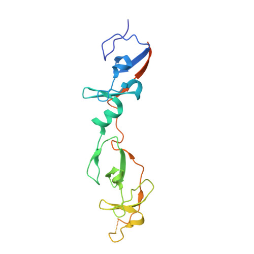
2DFY

Crystal structure of a cyclized protein fusion of LMO4 LIM domains 1 and 2 with the LIM interacting domain of LDB1


Experimental Data Snapshot

  • Method: X-RAY DIFFRACTION
  • Resolution: 1.65 Å
  • R-Value Free: 0.194 
  • R-Value Work: 0.167 
  • R-Value Observed: 0.168 

Starting Model: experimental
View more details

wwPDB Validation   3D Report Full Report


This is version 1.5 of the entry. See complete history


Literature

Stabilization of a binary protein complex by intein-mediated cyclization

Jeffries, C.M.J.Graham, S.C.Stokes, P.H.Collyer, C.A.Guss, J.M.Matthews, J.M.

(2006) Protein Sci 15: 2612-2618

  • DOI: https://doi.org/10.1110/ps.062377006
  • Primary Citation of Related Structures:  
    2DFY

  • PubMed Abstract: 

    The study of protein-protein interactions can be hampered by the instability of one or more of the protein complex components. In this study, we showed that intein-mediated cyclization can be used to engineer an artificial intramolecular cyclic protein complex between two interacting proteins: the largely unstable LIM-only protein 4 (LMO4) and an unstructured domain of LIM domain binding protein 1 (ldb1). The X-ray structure of the cyclic complex is identical to noncyclized versions of the complex. Chemical and thermal denaturation assays using intrinsic tryptophan fluorescence and dynamic light scattering were used to compare the relative stabilities of the cyclized complex, the intermolecular (or free) complex, and two linear versions of the intramolecular complex (in which the interacting domains of LMO4 and ldb1 were fused, via a flexible linker, in either orientation). In terms of resistance to denaturation, the cyclic complex is the most stable variant and the intermolecular complex is the least stable; however, the two linear intramolecular variants show significant differences in stability. These differences appear to be related to the relative contact order (the average distance in sequence between residues that make contacts within a structure) of key binding residues at the interface of the two proteins. Thus, the restriction of the more stable component of a complex may enhance stability to a greater extent than restraining less stable components.


  • Organizational Affiliation

    School of Molecular and Microbial Biosciences, University of Sydney, New South Wales 2006, Australia.


Macromolecules
Find similar proteins by:  (by identity cutoff)  |  3D Structure
Entity ID: 1
MoleculeChains Sequence LengthOrganismDetailsImage
fusion protein of LIM domain transcription factor LMO4 and LIM domain-binding protein 1A [auth X],
B [auth C]
195Mus musculusMutation(s): 2 
UniProt
Find proteins for P61969 (Mus musculus)
Explore P61969 
Go to UniProtKB:  P61969
Entity Groups  
Sequence Clusters30% Identity50% Identity70% Identity90% Identity95% Identity100% Identity
UniProt GroupP61969
Sequence Annotations
Expand
  • Reference Sequence
Small Molecules
Ligands 2 Unique
IDChains Name / Formula / InChI Key2D Diagram3D Interactions
GOL
Query on GOL

Download Ideal Coordinates CCD File 
G [auth X],
L [auth C]
GLYCEROL
C3 H8 O3
PEDCQBHIVMGVHV-UHFFFAOYSA-N
ZN
Query on ZN

Download Ideal Coordinates CCD File 
C [auth X]
D [auth X]
E [auth X]
F [auth X]
H [auth C]
C [auth X],
D [auth X],
E [auth X],
F [auth X],
H [auth C],
I [auth C],
J [auth C],
K [auth C]
ZINC ION
Zn
PTFCDOFLOPIGGS-UHFFFAOYSA-N
Experimental Data & Validation

Experimental Data

  • Method: X-RAY DIFFRACTION
  • Resolution: 1.65 Å
  • R-Value Free: 0.194 
  • R-Value Work: 0.167 
  • R-Value Observed: 0.168 
  • Space Group: P 3
Unit Cell:
Length ( Å )Angle ( ˚ )
a = 61.333α = 90
b = 61.333β = 90
c = 93.019γ = 120
Software Package:
Software NamePurpose
REFMACrefinement
PDB_EXTRACTdata extraction
DENZOdata reduction
SCALEPACKdata scaling
PHASERphasing

Structure Validation

View Full Validation Report


Entry History 

Deposition Data

Revision History  (Full details and data files)

  • Version 1.0: 2006-10-31
    Type: Initial release
  • Version 1.1: 2008-04-01
    Changes: Version format compliance
  • Version 1.2: 2011-07-13
    Changes: Non-polymer description, Version format compliance
  • Version 1.3: 2017-08-23
    Changes: Source and taxonomy
  • Version 1.4: 2021-11-10
    Changes: Database references, Derived calculations
  • Version 1.5: 2023-10-25
    Changes: Data collection, Refinement description