2DC3 | pdb_00002dc3

Crystal structure of human cytoglobin at 1.68 angstroms resolution


Experimental Data Snapshot

  • Method: X-RAY DIFFRACTION
  • Resolution: 1.68 Å
  • R-Value Free: 
    0.181 (Depositor), 0.190 (DCC) 
  • R-Value Work: 
    0.140 (Depositor), 0.150 (DCC) 
  • R-Value Observed: 
    0.142 (Depositor) 

Starting Model: experimental
View more details

wwPDB Validation   3D Report Full Report


Ligand Structure Quality Assessment 

Created with Raphaël 2.3.0Worse 01 BetterLigand structure goodness of fit to experimental dataBest fitted HEMClick on this verticalbar to view details

This is version 1.4 of the entry. See complete history


Literature

High-resolution structure of human cytoglobin: identification of extra N- and C-termini and a new dimerization mode.

Makino, M.Sugimoto, H.Sawai, H.Kawada, N.Yoshizato, K.Shiro, Y.

(2006) Acta Crystallogr D Biol Crystallogr 62: 671-677

  • DOI: https://doi.org/10.1107/S0907444906013813
  • Primary Citation of Related Structures:  
    2DC3

  • PubMed Abstract: 

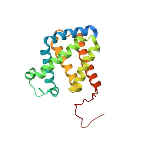
    Cytoglobin (Cgb) is a recently discovered member of the vertebrate haem-containing globin family. The structure of a new crystal form of wild-type human Cgb (space group C2) was determined at a resolution of 1.68 Angstrom. The results show the presence of an additional helix in the N-terminal residues (4-20) prior to the A helix and an ordered loop structure in the C-terminal region (168-188), while these extended peptides were invisible owing to disorder in the previously reported structures using a P3(2)21 crystal at a resolution of 2.4 Angstrom. A detailed comparison of the two crystal structures shows differences in the conformation of the residues (i.e. Arg84) in the haem environment owing to a different dimeric arrangement.


  • Organizational Affiliation

    Biometal Science Laboratory, Harima Institute, RIKEN SPring-8 Center, Harima Institute, Japan.


Macromolecules
Find similar proteins by:  (by identity cutoff)  |  3D Structure
Entity ID: 1
MoleculeChains Sequence LengthOrganismDetailsImage
Cytoglobin
A, B
193Homo sapiensMutation(s): 0 
Gene Names: CYGB
EC: 1.11.1 (UniProt), 1.14.12 (UniProt), 1.15.1.1 (UniProt), 1.7 (UniProt)
UniProt & NIH Common Fund Data Resources
Find proteins for Q8WWM9 (Homo sapiens)
Explore Q8WWM9 
Go to UniProtKB:  Q8WWM9
PHAROS:  Q8WWM9
GTEx:  ENSG00000161544 
Entity Groups  
Sequence Clusters30% Identity50% Identity70% Identity90% Identity95% Identity100% Identity
UniProt GroupQ8WWM9
Sequence Annotations
Expand
  • Reference Sequence
Experimental Data & Validation

Experimental Data

  • Method: X-RAY DIFFRACTION
  • Resolution: 1.68 Å
  • R-Value Free:  0.181 (Depositor), 0.190 (DCC) 
  • R-Value Work:  0.140 (Depositor), 0.150 (DCC) 
  • R-Value Observed: 0.142 (Depositor) 
Space Group: C 1 2 1
Unit Cell:
Length ( Å )Angle ( ˚ )
a = 78.53α = 90
b = 52.76β = 87.75
c = 83.44γ = 90
Software Package:
Software NamePurpose
REFMACrefinement
HKL-2000data reduction
SCALEPACKdata scaling
MOLREPphasing

Structure Validation

View Full Validation Report


Ligand Structure Quality Assessment 

Created with Raphaël 2.3.0Worse 01 BetterLigand structure goodness of fit to experimental dataBest fitted HEMClick on this verticalbar to view details

Entry History 

Deposition Data

Revision History  (Full details and data files)

  • Version 1.0: 2006-05-23
    Type: Initial release
  • Version 1.1: 2008-04-30
    Changes: Version format compliance
  • Version 1.2: 2011-07-13
    Changes: Version format compliance
  • Version 1.3: 2023-10-25
    Changes: Data collection, Database references, Derived calculations, Refinement description
  • Version 1.4: 2024-11-13
    Changes: Structure summary