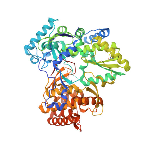
2D3U

X-ray crystal structure of hepatitis C virus RNA dependent RNA polymerase in complex with non-nucleoside analogue inhibitor


Experimental Data Snapshot

  • Method: X-RAY DIFFRACTION
  • Resolution: 2.00 Å
  • R-Value Free: 0.245 
  • R-Value Work: 0.208 
  • R-Value Observed: 0.208 

wwPDB Validation   3D Report Full Report


Ligand Structure Quality Assessment 


This is version 1.4 of the entry. See complete history


Literature

Non-nucleoside Inhibitors Binding to Hepatitis C Virus NS5B Polymerase Reveal a Novel Mechanism of Inhibition

Biswal, B.K.Wang, M.Cherney, M.M.Chan, L.Yannopoulos, C.G.Bilimoria, D.Bedard, J.James, M.N.G.

(2006) J Mol Biol 361: 33-45

  • DOI: https://doi.org/10.1016/j.jmb.2006.05.074
  • Primary Citation of Related Structures:  
    2D3U, 2D3Z, 2D41

  • PubMed Abstract: 

    The RNA-dependent RNA polymerase (NS5B) from hepatitis C virus (HCV) is a key enzyme in HCV replication. NS5B is a major target for the development of antiviral compounds directed against HCV. Here we present the structures of three thiophene-based non-nucleoside inhibitors (NNIs) bound non-covalently to NS5B. Each of the inhibitors binds to NS5B non-competitively to a common binding site in the "thumb" domain that is approximately 35 Angstroms from the polymerase active site located in the "palm" domain. The three compounds exhibit IC(50) values in the range of 270 nM to 307 nM and have common binding features that result in relatively large conformational changes of residues that interact directly with the inhibitors as well as for other residues adjacent to the binding site. Detailed comparisons of the unbound NS5B structure with those having the bound inhibitors present show that residues Pro495 to Arg505 (the N terminus of the "T" helix) exhibit some of the largest changes. It has been reported that Pro495, Pro496, Val499 and Arg503 are part of the guanosine triphosphate (GTP) specific allosteric binding site located in close proximity to our binding site. It has also been reported that the introduction of mutations to key residues in this region (i.e. Val499Gly) ablate in vivo sub-genomic HCV RNA replication. The details of NS5B polymerase/inhibitor binding interactions coupled with the observed induced conformational changes provide new insights into the design of novel NNIs of HCV.


  • Organizational Affiliation

    Canadian Institutes of Health Research (CIHR) Group in Protein Structure and Function, Department of Biochemistry, University of Alberta, Edmonton, Canada.


Macromolecules
Find similar proteins by:  (by identity cutoff)  |  3D Structure
Entity ID: 1
MoleculeChains Sequence LengthOrganismDetailsImage
polyprotein
A, B
570Hepacivirus hominisMutation(s): 0 
Gene Names: Type 1B
EC: 2.7.7.48
UniProt
Find proteins for Q99AU2 (Hepatitis C virus subtype 1b)
Explore Q99AU2 
Go to UniProtKB:  Q99AU2
Entity Groups  
Sequence Clusters30% Identity50% Identity70% Identity90% Identity95% Identity100% Identity
UniProt GroupQ99AU2
Sequence Annotations
Expand
  • Reference Sequence
Small Molecules
Ligands 1 Unique
IDChains Name / Formula / InChI Key2D Diagram3D Interactions
CCT
Query on CCT

Download Ideal Coordinates CCD File 
C [auth A],
D [auth B]
5-(4-CYANOPHENYL)-3-{[(2-METHYLPHENYL)SULFONYL]AMINO}THIOPHENE-2-CARBOXYLIC ACID
C19 H14 N2 O4 S2
RYXAHMSZLLIPRO-UHFFFAOYSA-N
Binding Affinity Annotations 
IDSourceBinding Affinity
CCT PDBBind:  2D3U Ki: 120 (nM) from 1 assay(s)
Experimental Data & Validation

Experimental Data

  • Method: X-RAY DIFFRACTION
  • Resolution: 2.00 Å
  • R-Value Free: 0.245 
  • R-Value Work: 0.208 
  • R-Value Observed: 0.208 
  • Space Group: P 21 21 21
Unit Cell:
Length ( Å )Angle ( ˚ )
a = 84.925α = 90
b = 104.314β = 90
c = 127.013γ = 90
Software Package:
Software NamePurpose
CNSrefinement
SCALEPACKdata scaling
CNSphasing

Structure Validation

View Full Validation Report


Ligand Structure Quality Assessment 


Entry History 

Deposition Data

Revision History  (Full details and data files)

  • Version 1.0: 2006-08-01
    Type: Initial release
  • Version 1.1: 2008-04-30
    Changes: Version format compliance
  • Version 1.2: 2011-07-13
    Changes: Version format compliance
  • Version 1.3: 2017-10-11
    Changes: Refinement description
  • Version 1.4: 2024-11-13
    Changes: Data collection, Database references, Derived calculations, Structure summary