2D31

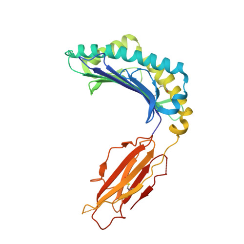
Crystal structure of disulfide-linked HLA-G dimer


Experimental Data Snapshot

  • Method: X-RAY DIFFRACTION
  • Resolution: 3.20 Å
  • R-Value Free: 0.298 
  • R-Value Work: 0.235 
  • R-Value Observed: 0.239 

Starting Model: experimental
View more details

wwPDB Validation   3D Report Full Report


This is version 1.6 of the entry. See complete history


Literature

Efficient Leukocyte Ig-like Receptor Signaling and Crystal Structure of Disulfide-linked HLA-G Dimer

Shiroishi, M.Kuroki, K.Ose, T.Rasubala, L.Shiratori, I.Arase, H.Tsumoto, K.Kumagai, I.Kohda, D.Maenaka, K.

(2006) J Biol Chem 281: 10439-10447

  • DOI: https://doi.org/10.1074/jbc.M512305200
  • Primary Citation of Related Structures:  
    2D31

  • PubMed Abstract: 

    HLA-G is a nonclassical major histocompatibility complex class I (MHCI) molecule, which is expressed in trophoblasts and confers immunological tolerance in the maternal-fetal interface by binding to leukocyte Ig-like receptors (LILRs, also called as LIR/ILT/CD85) and CD8. HLA-G is expressed in disulfide-linked dimer form both in solution and at the cell surface. Interestingly, MHCI dimer formations have been involved in pathogenesis and T cell activation. The structure and receptor binding characteristics of MHCI dimers have never been evaluated. Here we performed binding studies showing that the HLA-G dimer exhibited higher overall affinity to LILRB1/2 than the monomer by significant avidity effects. Furthermore, the cell reporter assay demonstrated that the dimer formation remarkably enhanced the LILRB1-mediated signaling at the cellular level. We further determined the crystal structure of the wild-type dimer of HLA-G with the intermolecular Cys(42)-Cys(42) disulfide bond. This dimer structure showed the oblique configuration to expose two LILR/CD8-binding sites upward from the membrane easily accessible for receptors, providing plausible 1:2 (HLA-G dimer:receptors) complex models. These results indicated that the HLA-G dimer conferred increased avidity in a proper structural orientation to induce efficient LILR signaling, resulting in the dominant immunosuppressive effects. Moreover, structural and functional implications for other MHCI dimers observed in activated T cells and the pathogenic allele, HLA-B27, are discussed.


  • Organizational Affiliation

    Division of Structural Biology, Medical Institute of Bioregulation, Kyushu University, 3-1-1 Maidashi, Higashi-ku, Fukuoka 812-8582, Japan.


Macromolecules
Find similar proteins by:  (by identity cutoff)  |  3D Structure
Entity ID: 1
MoleculeChains Sequence LengthOrganismDetailsImage
HLA class I histocompatibility antigen, alpha chain G
A, D
276Homo sapiensMutation(s): 0 
UniProt & NIH Common Fund Data Resources
Find proteins for P17693 (Homo sapiens)
Explore P17693 
Go to UniProtKB:  P17693
PHAROS:  P17693
GTEx:  ENSG00000204632 
Entity Groups  
Sequence Clusters30% Identity50% Identity70% Identity90% Identity95% Identity100% Identity
UniProt GroupP17693
Sequence Annotations
Expand
  • Reference Sequence
Find similar proteins by:  (by identity cutoff)  |  3D Structure
Entity ID: 2
MoleculeChains Sequence LengthOrganismDetailsImage
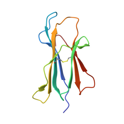
Beta-2-microglobulin
B, E
100Homo sapiensMutation(s): 0 
UniProt & NIH Common Fund Data Resources
Find proteins for P61769 (Homo sapiens)
Explore P61769 
Go to UniProtKB:  P61769
PHAROS:  P61769
GTEx:  ENSG00000166710 
Entity Groups  
Sequence Clusters30% Identity50% Identity70% Identity90% Identity95% Identity100% Identity
UniProt GroupP61769
Sequence Annotations
Expand
  • Reference Sequence

Find similar proteins by:  Sequence   |   3D Structure  

Entity ID: 3
MoleculeChains Sequence LengthOrganismDetailsImage
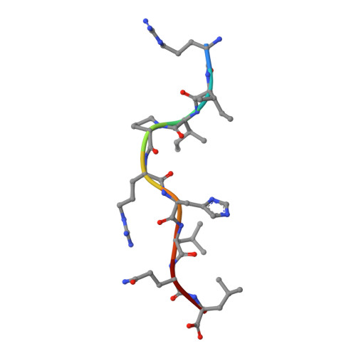
9-mer peptide from Histone H2A
C, F
9Homo sapiensMutation(s): 0 
UniProt & NIH Common Fund Data Resources
Find proteins for P16104 (Homo sapiens)
Explore P16104 
Go to UniProtKB:  P16104
PHAROS:  P16104
GTEx:  ENSG00000188486 
Entity Groups  
UniProt GroupP16104
Sequence Annotations
Expand
  • Reference Sequence
Experimental Data & Validation

Experimental Data

  • Method: X-RAY DIFFRACTION
  • Resolution: 3.20 Å
  • R-Value Free: 0.298 
  • R-Value Work: 0.235 
  • R-Value Observed: 0.239 
  • Space Group: P 21 21 2
Unit Cell:
Length ( Å )Angle ( ˚ )
a = 94.641α = 90
b = 127.807β = 90
c = 72.58γ = 90
Software Package:
Software NamePurpose
REFMACrefinement
HKL-2000data reduction
SCALEPACKdata scaling
MOLREPphasing

Structure Validation

View Full Validation Report


Entry History 

Deposition Data

Revision History  (Full details and data files)

  • Version 1.0: 2006-03-14
    Type: Initial release
  • Version 1.1: 2008-04-30
    Changes: Version format compliance
  • Version 1.2: 2011-07-13
    Changes: Version format compliance
  • Version 1.3: 2020-02-05
    Changes: Database references, Derived calculations
  • Version 1.4: 2020-02-19
    Changes: Data collection, Derived calculations, Source and taxonomy
  • Version 1.5: 2023-10-25
    Changes: Data collection, Database references, Refinement description
  • Version 1.6: 2024-11-20
    Changes: Structure summary