2CEJ | pdb_00002cej

P1' Extended HIV-1 Protease Inhibitors Encompassing a Tertiary Alcohol in the Transition-State Mimicking Scaffold


Experimental Data Snapshot

  • Method: X-RAY DIFFRACTION
  • Resolution: 2.50 Å
  • R-Value Free: 
    0.277 (Depositor), 0.221 (DCC) 
  • R-Value Work: 
    0.236 (Depositor), 0.222 (DCC) 
  • R-Value Observed: 
    0.236 (Depositor) 

Starting Model: experimental
View more details

wwPDB Validation 3D Report Full Report

Validation slider image for 2CEJ

Ligand Structure Quality Assessment 


This is version 1.3 of the entry. See complete history

Literature

Microwave-Accelerated Synthesis of P1'-Extended HIV-1 Protease Inhibitors Encompassing a Tertiary Alcohol in the Transition-State Mimicking Scaffold

Ekegren, J.K.Ginman, N.Johansson, A.Wallberg, H.Larhed, M.Samuelsson, B.Unge, T.Hallberg, A.

(2006) J Med Chem 49: 1828

  • DOI: https://doi.org/10.1021/jm051239z
  • Primary Citation Related Structures: 
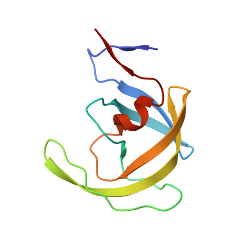
    2CEJ, 2CEM, 2CEN

  • PubMed Abstract: 

    Two series of P1'-extended HIV-1 protease inhibitors comprising a tertiary alcohol in the transition-state mimic exhibiting Ki values ranging from 2.1 to 93 nM have been synthesized. Microwave-accelerated palladium-catalyzed cross-couplings were utilized to rapidly optimize the P1' side chain. High cellular antiviral potencies were encountered when the P1' benzyl group was elongated with a 3- or 4-pyridyl substituent (EC50 = 0.18-0.22 microM). X-ray crystallographic data were obtained for three inhibitors cocrystallized with the enzyme.


  • Organizational Affiliation
    • Department of Medicinal Chemistry, Organic Pharmaceutical Chemistry, BMC, Uppsala University, Box 574, SE-751 23 Uppsala, Sweden.

Macromolecule Content 

  • Total Structure Weight: 22.29 kDa 
  • Atom Count: 1,642 
  • Modeled Residue Count: 198 
  • Deposited Residue Count: 198 
  • Unique protein chains: 1

Macromolecules

Find similar proteins by:|  3D Structure
Entity ID: 1
MoleculeChains  Sequence LengthOrganismDetailsImage
POL PROTEIN
A, B
99Human immunodeficiency virus 1Mutation(s): 0 
EC: 3.4.23.16
UniProt
Find proteins for Q8Q3H0 (Human immunodeficiency virus type 1)
Explore Q8Q3H0 
Go to UniProtKB:  Q8Q3H0
Entity Groups
Sequence Clusters30% Identity50% Identity70% Identity90% Identity95% Identity100% Identity
UniProt GroupQ8Q3H0
Sequence Annotations
Expand
Reference Sequence

Small Molecules

Ligands 1 Unique
IDChains Name / Formula / InChI Key2D Diagram3D Interactions
1AH

Query on 1AH


Download:Ideal Coordinates CCD File
C [auth B]3-AMINO-3-BENZYL-[4.3.0]BICYCLO-1,6-DIAZANONAN-2-ONE
C34 H41 Br N4 O6
JTIFBCHFPVQSEH-UNMGZEBXSA-N

Experimental Data & Validation

Experimental Data

  • Method: X-RAY DIFFRACTION
  • Resolution: 2.50 Å
  • R-Value Free:  0.277 (Depositor), 0.221 (DCC) 
  • R-Value Work:  0.236 (Depositor), 0.222 (DCC) 
  • R-Value Observed: 0.236 (Depositor) 
Space Group: P 21 21 2
Unit Cell:
Length ( Å )Angle ( ˚ )
a = 57.88α = 90
b = 85.71β = 90
c = 46.82γ = 90
Software Package:
Software NamePurpose
CNSrefinement
MOSFLMdata reduction
SCALAdata scaling
CNSphasing

Structure Validation

View Full Validation Report


Ligand Structure Quality Assessment 


Entry History 

Deposition Data

Revision History  (Full details and data files)

  • Version 1.0: 2007-02-13
    Type: Initial release
  • Version 1.1: 2011-05-08
    Changes: Version format compliance
  • Version 1.2: 2011-07-13
    Changes: Version format compliance
  • Version 1.3: 2023-12-13
    Changes: Data collection, Database references, Other, Refinement description