2CAZ

ESCRT-I core


Experimental Data Snapshot

  • Method: X-RAY DIFFRACTION
  • Resolution: 3.60 Å
  • R-Value Free: 0.357 
  • R-Value Work: 0.328 
  • R-Value Observed: 0.329 

wwPDB Validation   3D Report Full Report


This is version 1.4 of the entry. See complete history


Literature

Escrt-I Core and Escrt-II Glue Domain Structures Reveal Role for Glue in Linking to Escrt-I and Membranes.

Teo, H.Gill, D.J.Sun, J.Perisic, O.Veprintsev, D.B.Wallis, Y.Emr, S.D.Williams, R.L.

(2006) Cell 125: 99

  • DOI: https://doi.org/10.1016/j.cell.2006.01.047
  • Primary Citation of Related Structures:  
    2CAY, 2CAZ

  • PubMed Abstract: 

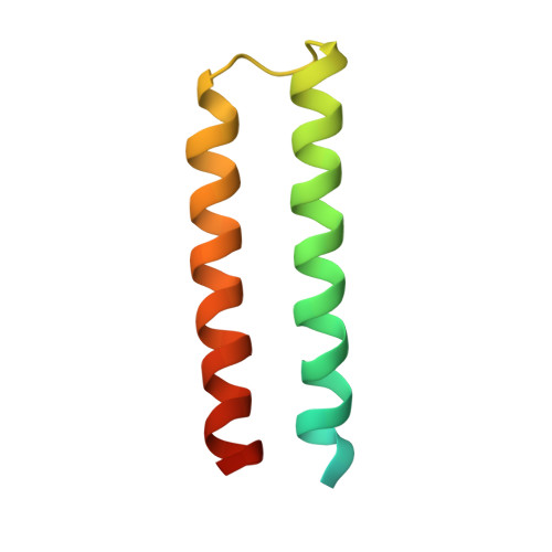
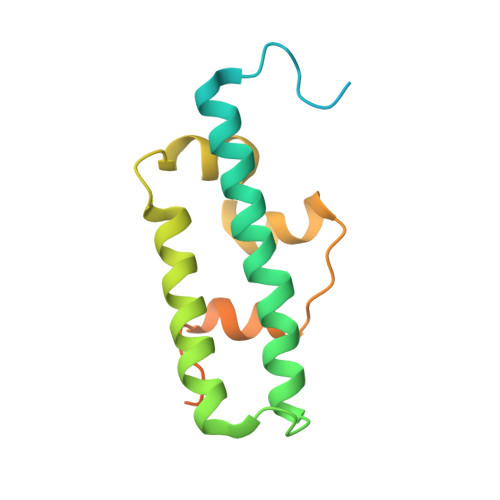
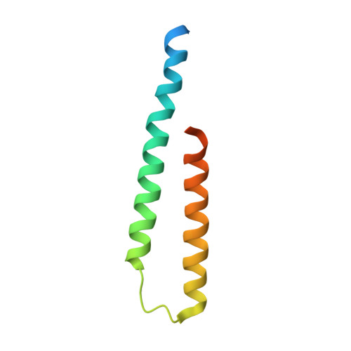
    ESCRT complexes form the main machinery driving protein sorting from endosomes to lysosomes. Currently, the picture regarding assembly of ESCRTs on endosomes is incomplete. The structure of the conserved heterotrimeric ESCRT-I core presented here shows a fan-like arrangement of three helical hairpins, each corresponding to a different subunit. Vps23/Tsg101 is the central hairpin sandwiched between the other subunits, explaining the critical role of its "steadiness box" in the stability of ESCRT-I. We show that yeast ESCRT-I links directly to ESCRT-II, through a tight interaction of Vps28 (ESCRT-I) with the yeast-specific zinc-finger insertion within the GLUE domain of Vps36 (ESCRT-II). The crystal structure of the GLUE domain missing this insertion reveals it is a split PH domain, with a noncanonical lipid binding pocket that binds PtdIns3P. The simultaneous and reinforcing interactions of ESCRT-II GLUE domain with membranes, ESCRT-I, and ubiquitin are critical for ubiquitinated cargo progression from early to late endosomes.


  • Organizational Affiliation

    MRC Laboratory of Molecular Biology, Medical Research Council Centre, Cambridge, CB2 2QH, United Kingdom. hlt@mrc-lmb.cam.ac.uk


Macromolecules
Find similar proteins by:  (by identity cutoff)  |  3D Structure
Entity ID: 1
MoleculeChains Sequence LengthOrganismDetailsImage
SUPPRESSOR PROTEIN STP22 OF TEMPERATURE-SENSITIVE ALPHA-FACTOR RECEPTOR AND ARGININE PERMEASE
A, D
82Saccharomyces cerevisiaeMutation(s): 0 
UniProt
Find proteins for P25604 (Saccharomyces cerevisiae (strain ATCC 204508 / S288c))
Explore P25604 
Go to UniProtKB:  P25604
Entity Groups  
Sequence Clusters30% Identity50% Identity70% Identity90% Identity95% Identity100% Identity
UniProt GroupP25604
Sequence Annotations
Expand
  • Reference Sequence
Find similar proteins by:  (by identity cutoff)  |  3D Structure
Entity ID: 2
MoleculeChains Sequence LengthOrganismDetailsImage
VACUOLAR PROTEIN SORTING-ASSOCIATED PROTEIN VPS28
B, E
155Saccharomyces cerevisiaeMutation(s): 0 
UniProt
Find proteins for Q02767 (Saccharomyces cerevisiae (strain ATCC 204508 / S288c))
Explore Q02767 
Go to UniProtKB:  Q02767
Entity Groups  
Sequence Clusters30% Identity50% Identity70% Identity90% Identity95% Identity100% Identity
UniProt GroupQ02767
Sequence Annotations
Expand
  • Reference Sequence
Find similar proteins by:  (by identity cutoff)  |  3D Structure
Entity ID: 3
MoleculeChains Sequence LengthOrganismDetailsImage
PROTEIN SRN2
C, F
85Saccharomyces cerevisiaeMutation(s): 0 
UniProt
Find proteins for Q99176 (Saccharomyces cerevisiae (strain ATCC 204508 / S288c))
Explore Q99176 
Go to UniProtKB:  Q99176
Entity Groups  
Sequence Clusters30% Identity50% Identity70% Identity90% Identity95% Identity100% Identity
UniProt GroupQ99176
Sequence Annotations
Expand
  • Reference Sequence
Experimental Data & Validation

Experimental Data

  • Method: X-RAY DIFFRACTION
  • Resolution: 3.60 Å
  • R-Value Free: 0.357 
  • R-Value Work: 0.328 
  • R-Value Observed: 0.329 
  • Space Group: P 61
Unit Cell:
Length ( Å )Angle ( ˚ )
a = 167.914α = 90
b = 167.914β = 90
c = 50.242γ = 120
Software Package:
Software NamePurpose
MOSFLMdata reduction
SCALAdata scaling
SnBphasing
SHARPphasing
REFMACrefinement

Structure Validation

View Full Validation Report


Entry History 

Deposition Data

Revision History  (Full details and data files)

  • Version 1.0: 2006-04-07
    Type: Initial release
  • Version 1.1: 2011-05-08
    Changes: Version format compliance
  • Version 1.2: 2011-07-13
    Changes: Version format compliance
  • Version 1.3: 2017-07-05
    Changes: Refinement description
  • Version 1.4: 2019-05-08
    Changes: Data collection, Experimental preparation