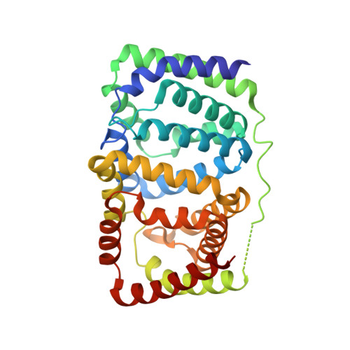
2C9E | pdb_00002c9e

Peridinin-chlorophyll a protein, high-salt form


Experimental Data Snapshot

  • Method: X-RAY DIFFRACTION
  • Resolution: 2.10 Å
  • R-Value Free: 
    0.219 (Depositor), 0.210 (DCC) 
  • R-Value Work: 
    0.174 (Depositor), 0.160 (DCC) 
  • R-Value Observed: 
    0.174 (Depositor) 

Starting Model: experimental
View more details

wwPDB Validation   3D Report Full Report


Ligand Structure Quality Assessment 

Created with Raphaël 2.3.0Worse 01 BetterLigand structure goodness of fit to experimental dataBest fitted DGDClick on this verticalbar to view detailsBest fitted CLAClick on this verticalbar to view detailsBest fitted PIDClick on this verticalbar to view details

This is version 2.0 of the entry. See complete history


Literature

X-Ray Structure of the High-Salt Form of the Peridinin-Chlorophyll A-Protein from the Dinoflagellate Amphidinium Carterae: Modulation of the Spectral Properties of Pigments by the Protein Environment.

Schulte, T.Sharples, F.P.Hiller, R.G.Hofmann, E.

(2009) Biochemistry 48: 4466

  • DOI: https://doi.org/10.1021/bi802320q
  • Primary Citation of Related Structures:  
    2C9E

  • PubMed Abstract: 

    Light-harvesting complexes have evolved into very different structures but fulfill the same function, efficient harvesting of solar energy. In these complexes, pigments are fine-tuned and properly arranged to gather incoming photons. In the photosynthetic dinoflagellate Amphidinium carterae, two variants of the soluble light-harvesting complex PCP have been found [main form PCP (MFPCP) and high-salt PCP (HSPCP)], which show small variations in their pigment arrangement and tuning mechanisms. This feature makes them ideal models for studying pigment-protein interactions. Here we present the X-ray structure of the monomeric HSPCP determined at 2.1 A resolution and compare it to the structure of trimeric MFPCP. Despite the high degree of structural similarity (rmsd C(alpha)-C(alpha) of 1.89 A), the sequence variations lead to a changed overall pigment composition which includes the loss of two carotenoid molecules and a dramatic rearrangement of the chlorophyll phytol chains and of internal lipid molecules. On the basis of a detailed structural comparison, we favor a macrocycle geometry distortion of the chlorophylls rather than an electrostatic effect to explain energetic splitting of the chlorophyll a Q(y) bands [Ilagan, R. P. (2006) Biochemistry 45, 14052-14063]. Our analysis supports their assignment of peridinin 611* as the single blue-shifted peridinin in HSPCP but also highlights another electrostatic feature due to glutamate 202 which could add to the observed binding site asymmetry of the 611*/621* peridinin pair.


  • Organizational Affiliation

    Biophysics, Department of Biology and Biotechnology, Ruhr-University Bochum, D-44780 Bochum, Germany.


Macromolecules
Find similar proteins by:  (by identity cutoff)  |  3D Structure
Entity ID: 1
MoleculeChains Sequence LengthOrganismDetailsImage
PERIDININ-CHLOROPHYLL A PROTEIN327Amphidinium carteraeMutation(s): 0 
UniProt
Find proteins for O76183 (Amphidinium carterae)
Explore O76183 
Go to UniProtKB:  O76183
Entity Groups  
Sequence Clusters30% Identity50% Identity70% Identity90% Identity95% Identity100% Identity
UniProt GroupO76183
Sequence Annotations
Expand
  • Reference Sequence
Small Molecules
Ligands 4 Unique
IDChains Name / Formula / InChI Key2D Diagram3D Interactions
DGD
Query on DGD

Download Ideal Coordinates CCD File 
G [auth A],
K [auth A]
DIGALACTOSYL DIACYL GLYCEROL (DGDG)
C51 H96 O15
LDQFLSUQYHBXSX-HXXRYREZSA-N
CLA
Query on CLA

Download Ideal Coordinates CCD File 
B [auth A],
C [auth A]
CHLOROPHYLL A
C55 H72 Mg N4 O5
ATNHDLDRLWWWCB-AENOIHSZSA-M
PID
Query on PID

Download Ideal Coordinates CCD File 
D [auth A]
E [auth A]
F [auth A]
H [auth A]
I [auth A]
PERIDININ
C39 H50 O7
UYRDHEJRPVSJFM-FROCQLDGSA-N
MG
Query on MG

Download Ideal Coordinates CCD File 
L [auth A]MAGNESIUM ION
Mg
JLVVSXFLKOJNIY-UHFFFAOYSA-N
Experimental Data & Validation

Experimental Data

  • Method: X-RAY DIFFRACTION
  • Resolution: 2.10 Å
  • R-Value Free:  0.219 (Depositor), 0.210 (DCC) 
  • R-Value Work:  0.174 (Depositor), 0.160 (DCC) 
  • R-Value Observed: 0.174 (Depositor) 
Space Group: P 1 21 1
Unit Cell:
Length ( Å )Angle ( ˚ )
a = 46.576α = 90
b = 62.673β = 100.89
c = 63.345γ = 90
Software Package:
Software NamePurpose
CNSrefinement
XDSdata reduction
XSCALEdata scaling
MOLREPphasing

Structure Validation

View Full Validation Report


Ligand Structure Quality Assessment 

Created with Raphaël 2.3.0Worse 01 BetterLigand structure goodness of fit to experimental dataBest fitted DGDClick on this verticalbar to view detailsBest fitted CLAClick on this verticalbar to view detailsBest fitted PIDClick on this verticalbar to view details

Entry History 

Deposition Data

Revision History  (Full details and data files)

  • Version 1.0: 2007-02-20
    Type: Initial release
  • Version 1.1: 2012-08-08
    Changes: Atomic model, Derived calculations, Non-polymer description, Version format compliance
  • Version 2.0: 2023-12-13
    Changes: Data collection, Database references, Derived calculations, Non-polymer description, Other, Refinement description