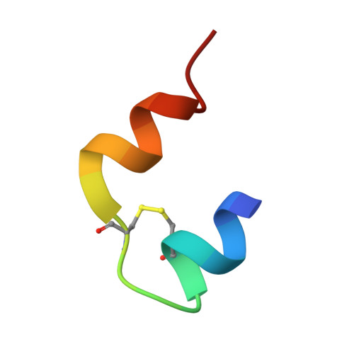
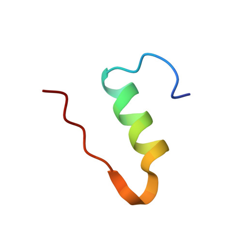
2C8Q

insuline(1sec) and UV laser excited fluorescence


Experimental Data Snapshot

  • Method: X-RAY DIFFRACTION
  • Resolution: 1.95 Å
  • R-Value Free: 0.230 
  • R-Value Work: 0.207 
  • R-Value Observed: 0.208 

wwPDB Validation   3D Report Full Report


This is version 1.2 of the entry. See complete history


Literature

Uv Laser-Excited Fluorescence as a Tool for the Visualization of Protein Crystals Mounted in Loops.

Vernede, X.Lavault, B.Ohana, J.Nurizzo, D.Joly, J.Jacquamet, L.Felisaz, F.Cipriani, F.Bourgeois, D.

(2006) Acta Crystallogr D Biol Crystallogr 62: 253

  • DOI: https://doi.org/10.1107/S0907444905041429
  • Primary Citation of Related Structures:  
    2C8O, 2C8P, 2C8Q, 2C8R

  • PubMed Abstract: 

    Structural proteomics has promoted the rapid development of automated protein structure determination using X-ray crystallography. Robotics are now routinely used along the pipeline from genes to protein structures. However, a bottleneck still remains. At synchrotron beamlines, the success rate of automated sample alignment along the X-ray beam is limited by difficulties in visualization of protein crystals, especially when they are small and embedded in mother liquor. Despite considerable improvement in optical microscopes, the use of visible light transmitted or reflected by the sample may result in poor or misleading contrast. Here, the endogenous fluorescence from aromatic amino acids has been used to identify even tiny or weakly fluorescent crystals with a high success rate. The use of a compact laser at 266 nm in combination with non-fluorescent sample holders provides an efficient solution to collect high-contrast fluorescence images in a few milliseconds and using standard camera optics. The best image quality was obtained with direct illumination through a viewing system coaxial with the UV beam. Crystallographic data suggest that the employed UV exposures do not generate detectable structural damage.


  • Organizational Affiliation

    LCCP, UMR 5075, IBS-CEA/CNRS/UJF, Grenoble, France.


Macromolecules

Find similar proteins by:  Sequence   |   3D Structure  

Entity ID: 1
MoleculeChains Sequence LengthOrganismDetailsImage
INSULIN A CHAIN21Homo sapiensMutation(s): 0 
UniProt & NIH Common Fund Data Resources
Find proteins for P01308 (Homo sapiens)
Explore P01308 
Go to UniProtKB:  P01308
PHAROS:  P01308
GTEx:  ENSG00000254647 
Entity Groups  
Sequence Clusters30% Identity50% Identity70% Identity90% Identity95% Identity100% Identity
UniProt GroupP01308
Sequence Annotations
Expand
  • Reference Sequence
Find similar proteins by:  (by identity cutoff)  |  3D Structure
Entity ID: 2
MoleculeChains Sequence LengthOrganismDetailsImage
INSULIN B CHAIN29Homo sapiensMutation(s): 0 
UniProt & NIH Common Fund Data Resources
Find proteins for P01308 (Homo sapiens)
Explore P01308 
Go to UniProtKB:  P01308
PHAROS:  P01308
GTEx:  ENSG00000254647 
Entity Groups  
Sequence Clusters30% Identity50% Identity70% Identity90% Identity95% Identity100% Identity
UniProt GroupP01308
Sequence Annotations
Expand
  • Reference Sequence
Experimental Data & Validation

Experimental Data

  • Method: X-RAY DIFFRACTION
  • Resolution: 1.95 Å
  • R-Value Free: 0.230 
  • R-Value Work: 0.207 
  • R-Value Observed: 0.208 
  • Space Group: I 21 3
Unit Cell:
Length ( Å )Angle ( ˚ )
a = 78.608α = 90
b = 78.608β = 90
c = 78.608γ = 90
Software Package:
Software NamePurpose
REFMACrefinement
MOSFLMdata reduction
SCALAdata scaling
AMoREphasing

Structure Validation

View Full Validation Report


Entry History 

Deposition Data

Revision History  (Full details and data files)

  • Version 1.0: 2006-03-08
    Type: Initial release
  • Version 1.1: 2011-07-13
    Changes: Advisory, Version format compliance
  • Version 1.2: 2023-12-13
    Changes: Data collection, Database references, Other, Refinement description