2C7N

Human Rabex-5 residues 1-74 in complex with Ubiquitin


Experimental Data Snapshot

  • Method: X-RAY DIFFRACTION
  • Resolution: 2.10 Å
  • R-Value Free: 0.238 
  • R-Value Work: 0.195 
  • R-Value Observed: 0.198 

wwPDB Validation   3D Report Full Report


This is version 1.2 of the entry. See complete history


Literature

Crystal Structure of the Ubiquitin Binding Domains of Rabex-5 Reveals Two Modes of Interaction with Ubiquitin.

Penengo, L.Mapelli, M.Murachelli, A.G.Confalonieri, S.Magri, L.Musacchio, A.Di Fiore, P.P.Polo, S.Schneider, T.R.

(2006) Cell 124: 1183

  • DOI: https://doi.org/10.1016/j.cell.2006.02.020
  • Primary Citation of Related Structures:  
    2C7M, 2C7N

  • PubMed Abstract: 

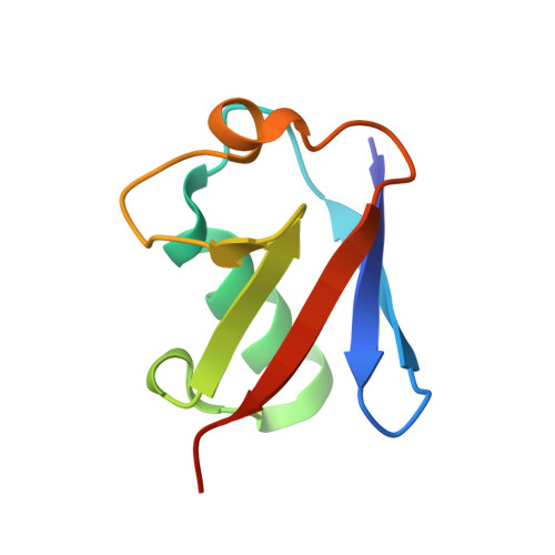
    The interaction between ubiquitinated proteins and intracellular proteins harboring ubiquitin binding domains (UBDs) is critical to a multitude of cellular processes. Here, we report that Rabex-5, a guanine nucleotide exchange factor for Rab5, binds to Ub through two independent UBDs. These UBDs determine a number of properties of Rabex-5, including its coupled monoubiquitination and interaction in vivo with ubiquitinated EGFRs. Structural and biochemical characterization of the UBDs of Rabex-5 revealed that one of them (MIU, motif interacting with ubiquitin) binds to Ub with modes superimposable to those of the UIM (ubiquitin-interacting motif):Ub interaction, although in the opposite orientation. The other UBD, RUZ (Rabex-5 ubiquitin binding zinc finger) binds to a surface of Ub centered on Asp58(Ub) and distinct from the "canonical" Ile44(Ub)-based surface. The two binding surfaces allow Ub to interact simultaneously with different UBDs, thus opening new perspectives in Ub-mediated signaling.


  • Organizational Affiliation

    IFOM, the FIRC Institute for Molecular Oncology Foundation, Via Adamello 16, 20139 Milan, Italy.


Macromolecules
Find similar proteins by:  (by identity cutoff)  |  3D Structure
Entity ID: 1
MoleculeChains Sequence LengthOrganismDetailsImage
RAB GUANINE NUCLEOTIDE EXCHANGE FACTOR 1
A, C, E, G, I
A, C, E, G, I, K
74Homo sapiensMutation(s): 0 
UniProt & NIH Common Fund Data Resources
Find proteins for Q9UJ41 (Homo sapiens)
Explore Q9UJ41 
Go to UniProtKB:  Q9UJ41
GTEx:  ENSG00000154710 
Entity Groups  
Sequence Clusters30% Identity50% Identity70% Identity90% Identity95% Identity100% Identity
UniProt GroupQ9UJ41
Sequence Annotations
Expand
  • Reference Sequence
Find similar proteins by:  (by identity cutoff)  |  3D Structure
Entity ID: 2
MoleculeChains Sequence LengthOrganismDetailsImage
UBIQUITIN
B, D, F, H, J
B, D, F, H, J, L
76Bos taurusMutation(s): 0 
UniProt
Find proteins for P0CH28 (Bos taurus)
Explore P0CH28 
Go to UniProtKB:  P0CH28
Entity Groups  
Sequence Clusters30% Identity50% Identity70% Identity90% Identity95% Identity100% Identity
UniProt GroupP0CH28
Sequence Annotations
Expand
  • Reference Sequence
Experimental Data & Validation

Experimental Data

  • Method: X-RAY DIFFRACTION
  • Resolution: 2.10 Å
  • R-Value Free: 0.238 
  • R-Value Work: 0.195 
  • R-Value Observed: 0.198 
  • Space Group: P 1
Unit Cell:
Length ( Å )Angle ( ˚ )
a = 44.3α = 108.2
b = 68.9β = 102.7
c = 98.5γ = 90.4
Software Package:
Software NamePurpose
REFMACrefinement
DENZOdata reduction
SCALEPACKdata scaling
HKL2MAPphasing

Structure Validation

View Full Validation Report


Entry History 

Deposition Data

Revision History  (Full details and data files)

  • Version 1.0: 2006-02-15
    Type: Initial release
  • Version 1.1: 2011-07-13
    Changes: Advisory, Refinement description, Version format compliance
  • Version 1.2: 2019-05-08
    Changes: Data collection, Experimental preparation, Other