2C6F

Structure of human somatic angiontensin-I converting enzyme N domain


Experimental Data Snapshot

  • Method: X-RAY DIFFRACTION
  • Resolution: 3.01 Å
  • R-Value Free: 0.273 
  • R-Value Work: 0.224 
  • R-Value Observed: 0.224 

wwPDB Validation   3D Report Full Report


Ligand Structure Quality Assessment 


This is version 2.1 of the entry. See complete history


Literature

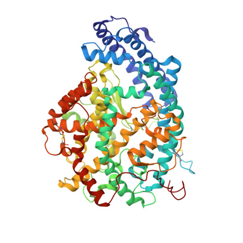
Crystal Structure of the N Domain of Human Somatic Angiotensin I-Converting Enzyme Provides a Structural Basis for Domain-Specific Inhibitor Design.

Corradi, H.R.Schwager, S.L.U.Nchinda, A.T.Sturrock, E.D.Acharya, K.R.

(2006) J Mol Biol 357: 964

  • DOI: https://doi.org/10.1016/j.jmb.2006.01.048
  • Primary Citation of Related Structures:  
    2C6F, 2C6N

  • PubMed Abstract: 

    Human somatic angiotensin I-converting enzyme (sACE) is a key regulator of blood pressure and an important drug target for combating cardiovascular and renal disease. sACE comprises two homologous metallopeptidase domains, N and C, joined by an inter-domain linker. Both domains are capable of cleaving the two hemoregulatory peptides angiotensin I and bradykinin, but differ in their affinities for a range of other substrates and inhibitors. Previously we determined the structure of testis ACE (C domain); here we present the crystal structure of the N domain of sACE (both in the presence and absence of the antihypertensive drug lisinopril) in order to aid the understanding of how these two domains differ in specificity and function. In addition, the structure of most of the inter-domain linker allows us to propose relative domain positions for sACE that may contribute to the domain cooperativity. The structure now provides a platform for the design of "domain-specific" second-generation ACE inhibitors.


  • Organizational Affiliation

    Department of Biology and Biochemistry, University of Bath, Claverton Down, Bath BA2 7AY, UK.


Macromolecules
Find similar proteins by:  (by identity cutoff)  |  3D Structure
Entity ID: 1
MoleculeChains Sequence LengthOrganismDetailsImage
ANGIOTENSIN-CONVERTING ENZYME, SOMATIC ISOFORM
A, B
612Homo sapiensMutation(s): 0 
EC: 3.4.15.1
UniProt & NIH Common Fund Data Resources
Find proteins for P12821 (Homo sapiens)
Explore P12821 
Go to UniProtKB:  P12821
PHAROS:  P12821
GTEx:  ENSG00000159640 
Entity Groups  
Sequence Clusters30% Identity50% Identity70% Identity90% Identity95% Identity100% Identity
UniProt GroupP12821
Sequence Annotations
Expand
  • Reference Sequence
Oligosaccharides

Help

Entity ID: 2
MoleculeChains Length2D Diagram Glycosylation3D Interactions
2-acetamido-2-deoxy-beta-D-glucopyranose-(1-4)-2-acetamido-2-deoxy-beta-D-glucopyranose
C, D, E, F
2N-Glycosylation
Glycosylation Resources
GlyTouCan:  G42666HT
GlyCosmos:  G42666HT
GlyGen:  G42666HT
Small Molecules
Ligands 5 Unique
IDChains Name / Formula / InChI Key2D Diagram3D Interactions
NAG
Query on NAG

Download Ideal Coordinates CCD File 
G [auth A],
H [auth A],
I [auth A],
O [auth B],
P [auth B]
2-acetamido-2-deoxy-beta-D-glucopyranose
C8 H15 N O6
OVRNDRQMDRJTHS-FMDGEEDCSA-N
GOL
Query on GOL

Download Ideal Coordinates CCD File 
N [auth A],
T [auth B]
GLYCEROL
C3 H8 O3
PEDCQBHIVMGVHV-UHFFFAOYSA-N
ZN
Query on ZN

Download Ideal Coordinates CCD File 
J [auth A],
Q [auth B]
ZINC ION
Zn
PTFCDOFLOPIGGS-UHFFFAOYSA-N
ACT
Query on ACT

Download Ideal Coordinates CCD File 
L [auth A],
M [auth A],
S [auth B]
ACETATE ION
C2 H3 O2
QTBSBXVTEAMEQO-UHFFFAOYSA-M
CL
Query on CL

Download Ideal Coordinates CCD File 
K [auth A],
R [auth B]
CHLORIDE ION
Cl
VEXZGXHMUGYJMC-UHFFFAOYSA-M
Experimental Data & Validation

Experimental Data

  • Method: X-RAY DIFFRACTION
  • Resolution: 3.01 Å
  • R-Value Free: 0.273 
  • R-Value Work: 0.224 
  • R-Value Observed: 0.224 
  • Space Group: C 2 2 21
Unit Cell:
Length ( Å )Angle ( ˚ )
a = 101.12α = 90
b = 211.32β = 90
c = 171.27γ = 90
Software Package:
Software NamePurpose
CNSrefinement
HKL-2000data reduction
HKL-2000data scaling
MOLREPphasing

Structure Validation

View Full Validation Report


Ligand Structure Quality Assessment 


Entry History 

Deposition Data

Revision History  (Full details and data files)

  • Version 1.0: 2006-11-08
    Type: Initial release
  • Version 1.1: 2011-05-08
    Changes: Version format compliance
  • Version 1.2: 2011-07-13
    Changes: Version format compliance
  • Version 2.0: 2020-07-29
    Type: Remediation
    Reason: Carbohydrate remediation
    Changes: Advisory, Atomic model, Data collection, Derived calculations, Other, Structure summary
  • Version 2.1: 2023-12-13
    Changes: Advisory, Data collection, Database references, Refinement description, Structure summary