2C3X

Structure of iodinated CBM25 from Bacillus halodurans amylase in complex with maltotetraose


Experimental Data Snapshot

  • Method: X-RAY DIFFRACTION
  • Resolution: 2.70 Å
  • R-Value Free: 0.283 
  • R-Value Work: 0.212 
  • R-Value Observed: 0.215 

wwPDB Validation   3D Report Full Report


This is version 2.0 of the entry. See complete history


Literature

A Structural and Functional Analysis of Alpha-Glucan Recognition by Family 25 and 26 Carbohydrate-Binding Modules Reveals a Conserved Mode of Starch Recognition

Boraston, A.B.Healey, M.Klassen, J.Ficko-Blean, E.Lammerts Van Bueren, A.Law, V.

(2006) J Biol Chem 281: 587

  • DOI: https://doi.org/10.1074/jbc.M509958200
  • Primary Citation of Related Structures:  
    2C3G, 2C3H, 2C3V, 2C3W, 2C3X

  • PubMed Abstract: 

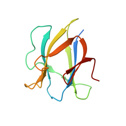
    Starch-hydrolyzing enzymes lacking alpha-glucan-specific carbohydrate-binding modules (CBMs) typically have lowered activity on granular starch relative to their counterparts with CBMs. Thus, consideration of starch recognition by CBMs is a key factor in understanding granular starch hydrolysis. To this end, we have dissected the modular structure of the maltohexaose-forming amylase from Bacillus halodurans (C-125). This five-module protein comprises an N-terminal family 13 catalytic module followed in order by two modules of unknown function, a family 26 CBM (BhCBM26), and a family 25 CBM (BhCBM25). Here we present a comprehensive structure-function analysis of starch and alpha-glucooligosaccharide recognition by BhCBM25 and BhCBM26 using UV methods, isothermal titration calorimetry, and x-ray crystallography. The results reveal that the two CBMs bind alpha-glucooligosaccharides, particularly those containing alpha-1,6 linkages, with different affinities but have similar abilities to bind granular starch. Notably, these CBMs appear to recognize the same binding sites in granular starch. The enhanced affinity of the tandem CBMs for granular starch is suggested to be the main biological advantage for this enzyme to contain two CBMs. Structural studies of the native and ligand-bound forms of BhCBM25 and BhCBM26 show a structurally conserved mode of ligand recognition but through non-sequence-conserved residues. Comparison of these CBM structures with other starch-specific CBM structures reveals a generally conserved mode of starch recognition.


  • Organizational Affiliation

    Biochemistry and Microbiology, University of Victoria, Victoria, British Columbia V8W 3P6, Canada. boraston@uvic.ca


Macromolecules
Find similar proteins by:  (by identity cutoff)  |  3D Structure
Entity ID: 1
MoleculeChains Sequence LengthOrganismDetailsImage
ALPHA-AMYLASE G-6
A, B
102Halalkalibacterium halodurans C-125Mutation(s): 0 
UniProt
Find proteins for Q9KFR4 (Halalkalibacterium halodurans (strain ATCC BAA-125 / DSM 18197 / FERM 7344 / JCM 9153 / C-125))
Explore Q9KFR4 
Go to UniProtKB:  Q9KFR4
Entity Groups  
Sequence Clusters30% Identity50% Identity70% Identity90% Identity95% Identity100% Identity
UniProt GroupQ9KFR4
Sequence Annotations
Expand
  • Reference Sequence
Oligosaccharides

Help

Entity ID: 2
MoleculeChains Length2D Diagram Glycosylation3D Interactions
alpha-D-glucopyranose-(1-4)-alpha-D-glucopyranose-(1-4)-alpha-D-glucopyranose-(1-4)-alpha-D-glucopyranose-(1-4)-alpha-D-glucopyranose
C
5N/A
Glycosylation Resources
GlyTouCan:  G50146AM
GlyCosmos:  G50146AM
Small Molecules
Ligands 1 Unique
IDChains Name / Formula / InChI Key2D Diagram3D Interactions
SO4
Query on SO4

Download Ideal Coordinates CCD File 
D [auth A]SULFATE ION
O4 S
QAOWNCQODCNURD-UHFFFAOYSA-L
Biologically Interesting Molecules (External Reference) 1 Unique
Experimental Data & Validation

Experimental Data

  • Method: X-RAY DIFFRACTION
  • Resolution: 2.70 Å
  • R-Value Free: 0.283 
  • R-Value Work: 0.212 
  • R-Value Observed: 0.215 
  • Space Group: P 43 21 2
Unit Cell:
Length ( Å )Angle ( ˚ )
a = 64.743α = 90
b = 64.743β = 90
c = 187.18γ = 90
Software Package:
Software NamePurpose
REFMACrefinement

Structure Validation

View Full Validation Report


Entry History 

Deposition Data

Revision History  (Full details and data files)

  • Version 1.0: 2005-10-17
    Type: Initial release
  • Version 1.1: 2011-05-07
    Changes: Version format compliance
  • Version 1.2: 2011-07-13
    Changes: Version format compliance
  • Version 2.0: 2020-07-29
    Type: Remediation
    Reason: Carbohydrate remediation
    Changes: Atomic model, Data collection, Derived calculations, Other, Structure summary