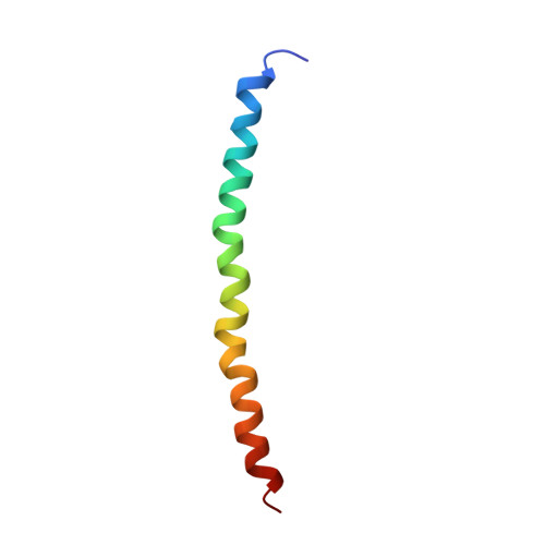
2C0W

Molecular Structure of fd Filamentous Bacteriophage Refined with Respect to X-ray Fibre Diffraction


Experimental Data Snapshot

  • Method: FIBER DIFFRACTION
  • Resolution: 3.20 Å
  • R-Value Work: 0.210 
  • R-Value Observed: 0.210 

Starting Model: experimental
View more details

wwPDB Validation   3D Report Full Report


This is version 1.3 of the entry. See complete history


Literature

Molecular Structure of Fd (F1, M13) Filamentous Bacteriophage Refined with Respect to X-Ray Fibre Diffraction and Solid-State NMR Data Supports Specific Models of Phage Assembly at the Bacterial Membrane.

Marvin, D.A.Welsh, L.C.Symmons, M.F.Scott, W.R.P.Straus, S.K.

(2006) J Mol Biol 355: 294

  • DOI: https://doi.org/10.1016/j.jmb.2005.10.048
  • Primary Citation of Related Structures:  
    2C0W, 2C0X

  • PubMed Abstract: 

    Filamentous bacteriophage (Inovirus) is a simple and well-characterized model system. The phage particle, or virion, is about 60 angstroms in diameter and several thousand angstrom units long. The virions are assembled at the bacterial membrane as they extrude out of the host without killing it, an example of specific transport of nucleoprotein assemblages across membranes. The Ff group (fd, f1 and M13) has been especially widely studied. Models of virion assembly have been proposed based on a molecular model of the fd virion derived by X-ray fibre diffraction. A somewhat different model of the fd virion using solid-state NMR data has been proposed, not consistent with these models of assembly nor with the X-ray diffraction data. Here we show that reinterpreted NMR data are also consistent with the model derived from X-ray fibre diffraction studies, and discuss models of virion assembly.


  • Organizational Affiliation

    Department of Biochemistry, University of Cambridge, Cambridge CB2 1GA, UK. d.a.marvin@bioc.cam.ac.uk


Macromolecules
Find similar proteins by:  (by identity cutoff)  |  3D Structure
Entity ID: 1
MoleculeChains Sequence LengthOrganismDetailsImage
COAT PROTEIN B50Enterobacteria phage fdMutation(s): 1 
UniProt
Find proteins for P69539 (Enterobacteria phage fd)
Explore P69539 
Go to UniProtKB:  P69539
Entity Groups  
Sequence Clusters30% Identity50% Identity70% Identity90% Identity95% Identity100% Identity
UniProt GroupP69539
Sequence Annotations
Expand
  • Reference Sequence
Experimental Data & Validation

Experimental Data

  • Method: FIBER DIFFRACTION
  • Resolution: 3.20 Å
  • R-Value Work: 0.210 
  • R-Value Observed: 0.210 
  • Space Group: P 1
Unit Cell:
Length ( Å )Angle ( ˚ )
a = 1α = 90
b = 1β = 90
c = 1γ = 90
Software Package:
Software NamePurpose
X-PLORrefinement
CCP13data reduction
CCP13-FDSCALEdata scaling
FX-PLORphasing

Structure Validation

View Full Validation Report


Entry History 

Deposition Data

Revision History  (Full details and data files)

  • Version 1.0: 2005-12-14
    Type: Initial release
  • Version 1.1: 2011-07-13
    Changes: Source and taxonomy, Version format compliance
  • Version 1.2: 2013-05-01
    Changes: Data collection, Source and taxonomy, Structure summary
  • Version 1.3: 2024-02-14
    Changes: Data collection, Database references, Derived calculations, Other, Refinement description