2C0L | pdb_00002c0l

TPR DOMAIN OF HUMAN PEX5P IN COMPLEX WITH HUMAN MSCP2


Experimental Data Snapshot

  • Method: X-RAY DIFFRACTION
  • Resolution: 2.30 Å
  • R-Value Free: 
    0.256 (Depositor), 0.288 (DCC) 
  • R-Value Work: 
    0.202 (Depositor), 0.222 (DCC) 
  • R-Value Observed: 
    0.205 (Depositor) 

wwPDB Validation 3D Report Full Report

Validation slider image for 2C0L

This is version 1.4 of the entry. See complete history

Literature

Recognition of a Functional Peroxisome Type 1 Target by the Dynamic Import Receptor Pex5P.

Stanley, W.A.Filipp, F.V.Kursula, P.Schuller, N.Erdmann, R.Schliebs, W.Sattler, M.Wilmanns, M.

(2006) Mol Cell 24: 653

  • DOI: https://doi.org/10.1016/j.molcel.2006.10.024
  • Primary Citation Related Structures: 
    2C0L, 2C0M

  • PubMed Abstract: 

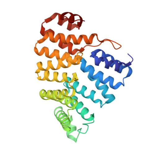
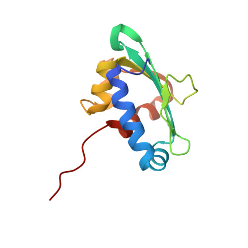
    Peroxisomes require the translocation of folded and functional target proteins of various sizes across the peroxisomal membrane. We have investigated the structure and function of the principal import receptor Pex5p, which recognizes targets bearing a C-terminal peroxisomal targeting signal type 1. Crystal structures of the receptor in the presence and absence of a peroxisomal target, sterol carrier protein 2, reveal major structural changes from an open, snail-like conformation into a closed, circular conformation. These changes are caused by a long loop C terminal to the 7-fold tetratricopeptide repeat segments. Mutations in residues of this loop lead to defects in peroxisomal import in human fibroblasts. The structure of the receptor/cargo complex demonstrates that the primary receptor-binding site of the cargo is structurally and topologically autonomous, enabling the cargo to retain its native structure and function.


  • Organizational Affiliation
    • European Molecular Biology Laboratory-Hamburg Outstation, Notkestrasse 85, 22603 Hamburg.

Macromolecule Content 

  • Total Structure Weight: 46.7 kDa 
  • Atom Count: 3,308 
  • Modeled Residue Count: 414 
  • Deposited Residue Count: 427 
  • Unique protein chains: 2

Macromolecules

Find similar proteins by:|  3D Structure
Entity ID: 1
MoleculeChains  Sequence LengthOrganismDetailsImage
PEROXISOMAL TARGETING SIGNAL 1 RECEPTOR305Homo sapiensMutation(s): 0 
UniProt & NIH Common Fund Data Resources
Find proteins for P50542 (Homo sapiens)
Explore P50542 
Go to UniProtKB:  P50542
PHAROS:  P50542
GTEx:  ENSG00000139197 
Entity Groups
Sequence Clusters30% Identity50% Identity70% Identity90% Identity95% Identity100% Identity
UniProt GroupP50542
Sequence Annotations
Expand
Reference Sequence
Find similar proteins by:|  3D Structure
Entity ID: 2
MoleculeChains  Sequence LengthOrganismDetailsImage
NONSPECIFIC LIPID-TRANSFER PROTEIN122Homo sapiensMutation(s): 0 
EC: 2.3.1.176 (UniProt), 2.3.1.155 (UniProt), 2.3.1.16 (UniProt)
UniProt & NIH Common Fund Data Resources
Find proteins for P22307 (Homo sapiens)
Explore P22307 
Go to UniProtKB:  P22307
PHAROS:  P22307
GTEx:  ENSG00000116171 
Entity Groups
Sequence Clusters30% Identity50% Identity70% Identity90% Identity95% Identity100% Identity
UniProt GroupP22307
Sequence Annotations
Expand
Reference Sequence

Experimental Data & Validation

Experimental Data

  • Method: X-RAY DIFFRACTION
  • Resolution: 2.30 Å
  • R-Value Free:  0.256 (Depositor), 0.288 (DCC) 
  • R-Value Work:  0.202 (Depositor), 0.222 (DCC) 
  • R-Value Observed: 0.205 (Depositor) 
Space Group: P 21 21 21
Unit Cell:
Length ( Å )Angle ( ˚ )
a = 40.48α = 90
b = 68.62β = 90
c = 137.39γ = 90
Software Package:
Software NamePurpose
REFMACrefinement
XDSdata reduction
XDSdata scaling
MOLREPphasing

Structure Validation

View Full Validation Report


Entry History 

Deposition Data

Revision History  (Full details and data files)

  • Version 1.0: 2006-11-15
    Type: Initial release
  • Version 1.1: 2011-05-08
    Changes: Version format compliance
  • Version 1.2: 2011-07-13
    Changes: Version format compliance
  • Version 1.3: 2019-07-24
    Changes: Data collection
  • Version 1.4: 2024-05-08
    Changes: Data collection, Database references, Other