Protonation of a Histidine Copper Ligand in Fern Plastocyanin.
Hulsker, R., Mery, A., Thomassen, E.A.J., Ranieri, A., Sola, M., Verbeet, M.P., Kohzuma, T., Ubbink, M.(2007) J Am Chem Soc 129: 4423
- PubMed: 17367139
- DOI: https://doi.org/10.1021/ja0690464
- Primary Citation of Related Structures:
2BZ7, 2BZC - PubMed Abstract:
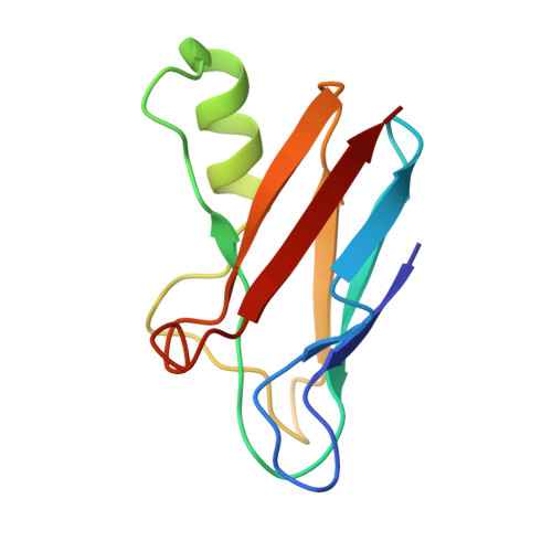
Plastocyanin is a small blue copper protein that shuttles electrons as part of the photosynthetic redox chain. Its redox behavior is changed at low pH as a result of protonation of the solvent-exposed copper-coordinating histidine. Protonation and subsequent redox inactivation could have a role in the down regulation of photosynthesis. As opposed to plastocyanin from other sources, in fern plastocyanin His90 protonation at low pH has been reported not to occur. Two possible reasons for that have been proposed: pi-pi stacking between Phe12 and His90 and lack of a hydrogen bond with the backbone oxygen of Gly36. We have produced this fern plastocyanin recombinantly and examined the properties of wild-type protein and mutants Phe12Leu, Gly36Pro, and the double mutant with NMR spectroscopy, X-ray crystallography, and cyclic voltammetry. The results demonstrate that, contrary to earlier reports, protonation of His90 in the wild-type protein does occur in solution with a pKa of 4.4 (+/-0.1). Neither the single mutants nor the double mutant exhibit a change in protonation behavior, indicating that the suggested interactions have no influence. The crystal structure at low pH of the Gly36Pro variant does not show His90 protonation, similar to what was found for the wild-type protein. The structure suggests that movement of the imidazole ring is hindered by crystal contacts. This study illustrates a significant difference between results obtained in solution by NMR and by crystallography.
- Leiden Institute of Chemistry, Leiden University, Gorlaeus Laboratories, P.O. Box 9502, 2300 RA, Leiden, The Netherlands.
Organizational Affiliation:


