2BV8 | pdb_00002bv8

The crystal structure of Phycocyanin from Gracilaria chilensis.


Experimental Data Snapshot

  • Method: X-RAY DIFFRACTION
  • Resolution: 2.01 Å
  • R-Value Free: 
    0.231 (Depositor), 0.219 (DCC) 
  • R-Value Work: 
    0.199 (Depositor), 0.191 (DCC) 
  • R-Value Observed: 
    0.199 (Depositor) 

Starting Model: experimental
View more details

wwPDB Validation 3D Report Full Report

Validation slider image for 2BV8

Ligand Structure Quality Assessment 


This is version 1.4 of the entry. See complete history

Literature

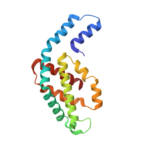
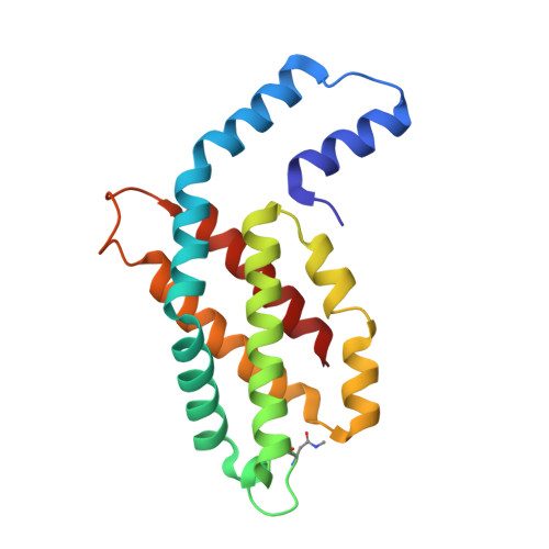
The Structure at 2 A Resolution of Phycocyanin from Gracilaria Chilensis and the Energy Transfer Network in a Pc-Pc Complex.

Contreras-Martel, C.Matamala, A.Bruna, C.Poo-Caama, G.Almonacid, D.Figueroa, M.Martinez-Oyanedel, J.Bunster, M.

(2007) Biophys Chem 125: 388

  • DOI: https://doi.org/10.1016/j.bpc.2006.09.014
  • Primary Citation Related Structures: 
    2BV8

  • PubMed Abstract: 

    Phycocyanin is a phycobiliprotein involved in light harvesting and conduction of light to the reaction centers in cyanobacteria and red algae. The structure of C-phycocyanin from Gracilaria chilensis was solved by X-ray crystallography at 2.0 A resolution in space group P2(1). An interaction model between two PC heterohexamers was built, followed by molecular dynamic refinement. The best model showed an inter-hexamer rotation of 23 degrees . The coordinates of a PC heterohexamer (alphabeta)(6) and of the PC-PC complex were used to perform energy transfer calculations between chromophores pairs using the fluorescence resonance energy transfer approach (FRET). Two main intra PC ((I)beta(3)(82)-->(I)alpha(1)(84)-->(I)alpha(5)(84)-->(I)beta(6)(82) and (I)beta(3)(153)-->(I)beta(5)(153)) and two main inter PC ((I)beta(6)(82)-->(II)beta(3)(82) and (I)beta(5)(153)-->(II)beta(3)(153)) pathways were proposed based on the values of the energy transfer constants calculated for all the chromophore pairs in the hexamer and in the complex.


  • Organizational Affiliation
    • Institut de Biologie Structurale, CEA-CNRS-UJF, Grenoble, France.

Macromolecule Content 

  • Total Structure Weight: 225.29 kDa 
  • Atom Count: 16,812 
  • Modeled Residue Count: 2,004 
  • Deposited Residue Count: 2,004 
  • Unique protein chains: 2

Macromolecules

Find similar proteins by:|  3D Structure
Entity ID: 1
MoleculeChains  Sequence LengthOrganismDetailsImage
C-PHYCOCYANIN ALPHA SUBUNIT162Agarophyton chilenseMutation(s): 0 
UniProt
Find proteins for Q6B8L6 (Gracilaria tenuistipitata var. liui)
Explore Q6B8L6 
Go to UniProtKB:  Q6B8L6
Entity Groups
Sequence Clusters30% Identity50% Identity70% Identity90% Identity95% Identity100% Identity
UniProt GroupQ6B8L6
Sequence Annotations
Expand
Reference Sequence
Find similar proteins by:|  3D Structure
Entity ID: 2
MoleculeChains  Sequence LengthOrganismDetailsImage
C-PHYCOCYANIN BETA SUBUNIT172Agarophyton chilenseMutation(s): 0 
UniProt
Find proteins for Q6B8L7 (Gracilaria tenuistipitata var. liui)
Explore Q6B8L7 
Go to UniProtKB:  Q6B8L7
Entity Groups
Sequence Clusters30% Identity50% Identity70% Identity90% Identity95% Identity100% Identity
UniProt GroupQ6B8L7
Sequence Annotations
Expand
Reference Sequence

Small Molecules

Ligands 2 Unique
IDChains Name / Formula / InChI Key2D Diagram3D Interactions
PEB

Query on PEB


Download:Ideal Coordinates CCD File
O [auth B]PHYCOERYTHROBILIN
C33 H40 N4 O6
NKCBCVIFPXGHAV-WAVSMFBNSA-N
CYC

Query on CYC


Download:Ideal Coordinates CCD File
AA [auth N]
BA [auth O]
CA [auth P]
DA [auth P]
M [auth A]
AA [auth N],
BA [auth O],
CA [auth P],
DA [auth P],
M [auth A],
N [auth B],
P [auth C],
Q [auth D],
R [auth D],
S [auth E],
T [auth F],
U [auth F],
V [auth K],
W [auth L],
X [auth L],
Y [auth M],
Z [auth N]
PHYCOCYANOBILIN
C33 H40 N4 O6
VXTXPYZGDQPMHK-GMXXPEQVSA-N
Modified Residues  1 Unique
IDChains TypeFormula2D DiagramParent
MEN
Query on MEN
B
D
F
H [auth L]
J [auth N]
B,
D,
F,
H [auth L],
J [auth N],
L [auth P]
L-PEPTIDE LINKINGC5 H10 N2 O3ASN

Experimental Data & Validation

Experimental Data

  • Method: X-RAY DIFFRACTION
  • Resolution: 2.01 Å
  • R-Value Free:  0.231 (Depositor), 0.219 (DCC) 
  • R-Value Work:  0.199 (Depositor), 0.191 (DCC) 
  • R-Value Observed: 0.199 (Depositor) 
Space Group: P 1 21 1
Unit Cell:
Length ( Å )Angle ( ˚ )
a = 101.985α = 90
b = 151.803β = 117.45
c = 101.548γ = 90
Software Package:
Software NamePurpose
CNSrefinement
XDSdata reduction
XSCALEdata scaling
CNSphasing

Structure Validation

View Full Validation Report


Ligand Structure Quality Assessment 


Entry History 

Deposition Data

Revision History  (Full details and data files)

  • Version 1.0: 2006-08-16
    Type: Initial release
  • Version 1.1: 2011-05-08
    Changes: Version format compliance
  • Version 1.2: 2011-07-13
    Changes: Version format compliance
  • Version 1.3: 2019-01-30
    Changes: Data collection, Experimental preparation, Other
  • Version 1.4: 2023-12-13
    Changes: Data collection, Database references, Derived calculations, Other, Refinement description