2BTF | pdb_00002btf

THE STRUCTURE OF CRYSTALLINE PROFILIN-BETA-ACTIN


Experimental Data Snapshot

  • Method: X-RAY DIFFRACTION
  • Resolution: 2.55 Å
  • R-Value Work: 
    0.199 (Depositor) 
  • R-Value Observed: 
    0.199 (Depositor) 

wwPDB Validation 3D Report Full Report

Validation slider image for 2BTF

This is version 1.4 of the entry. See complete history

Literature

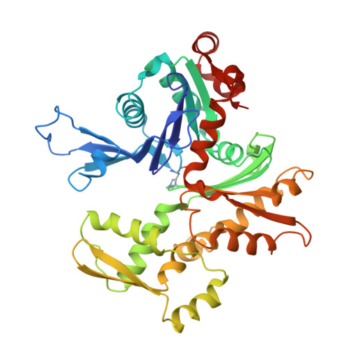
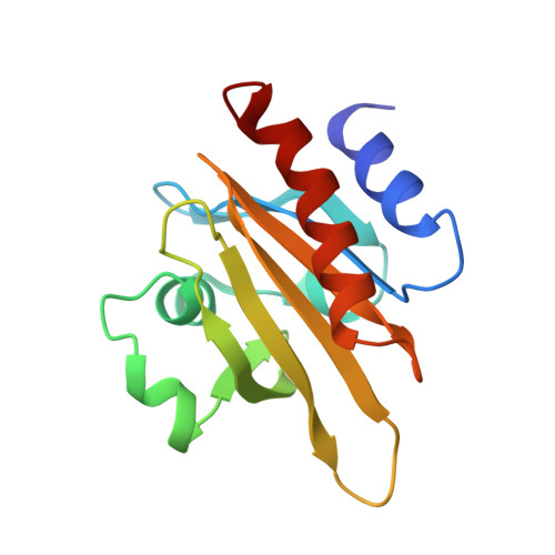
The structure of crystalline profilin-beta-actin.

Schutt, C.E.Myslik, J.C.Rozycki, M.D.Goonesekere, N.C.Lindberg, U.

(1993) Nature 365: 810-816

  • DOI: https://doi.org/10.1038/365810a0
  • Primary Citation Related Structures: 
    2BTF

  • PubMed Abstract: 

    The three-dimensional structure of bovine profilin-beta-actin has been solved to 2.55 A resolution by X-ray crystallography. There are several significant local changes in the structure of beta-actin compared with alpha-actin as well as an overall 5 degrees rotation between its two major domains. Actin molecules in the crystal are organized into ribbons through intermolecular contacts like those found in oligomeric protein assemblies. Profilin forms two extensive contacts with the actin ribbon, one of which appears to correspond to the solution contact in vitro.


  • Organizational Affiliation
    • Department of Chemistry, Henry H. Hoyt Laboratory, Princeton University, New Jersey 08544.

Macromolecule Content 

  • Total Structure Weight: 57.25 kDa 
  • Atom Count: 4,000 
  • Modeled Residue Count: 515 
  • Deposited Residue Count: 515 
  • Unique protein chains: 2

Macromolecules

Find similar proteins by:|  3D Structure
Entity ID: 1
MoleculeChains  Sequence LengthOrganismDetailsImage
BETA-ACTIN375Bos taurusMutation(s): 0 
EC: 3.6.4
UniProt
Find proteins for P60712 (Bos taurus)
Explore P60712 
Go to UniProtKB:  P60712
Entity Groups
Sequence Clusters30% Identity50% Identity70% Identity90% Identity95% Identity100% Identity
UniProt GroupP60712
Sequence Annotations
Expand
Reference Sequence
Find similar proteins by:|  3D Structure
Entity ID: 2
MoleculeChains  Sequence LengthOrganismDetailsImage
PROFILINB [auth P]140Bos taurusMutation(s): 0 
UniProt
Find proteins for P02584 (Bos taurus)
Explore P02584 
Go to UniProtKB:  P02584
Entity Groups
Sequence Clusters30% Identity50% Identity70% Identity90% Identity95% Identity100% Identity
UniProt GroupP02584
Sequence Annotations
Expand
Reference Sequence

Small Molecules

Ligands 2 Unique
IDChains Name / Formula / InChI Key2D Diagram3D Interactions
ATP

Query on ATP


Download:Ideal Coordinates CCD File
D [auth A]ADENOSINE-5'-TRIPHOSPHATE
C10 H16 N5 O13 P3
ZKHQWZAMYRWXGA-KQYNXXCUSA-N
SR

Query on SR


Download:Ideal Coordinates CCD File
C [auth A]STRONTIUM ION
Sr
PWYYWQHXAPXYMF-UHFFFAOYSA-N
Modified Residues  1 Unique
IDChains TypeFormula2D DiagramParent
HIC
Query on HIC
A
L-PEPTIDE LINKINGC7 H11 N3 O2HIS

Experimental Data & Validation

Experimental Data

  • Method: X-RAY DIFFRACTION
  • Resolution: 2.55 Å
  • R-Value Work:  0.199 (Depositor) 
  • R-Value Observed: 0.199 (Depositor) 
Space Group: P 21 21 21
Unit Cell:
Length ( Å )Angle ( ˚ )
a = 38.95α = 90
b = 71.3β = 90
c = 171.9γ = 90
Software Package:
Software NamePurpose
X-PLORmodel building
X-PLORrefinement
X-PLORphasing

Structure Validation

View Full Validation Report


Entry History 

Deposition Data

Revision History  (Full details and data files)

  • Version 1.0: 1994-06-22
    Type: Initial release
  • Version 1.1: 2008-03-03
    Changes: Version format compliance
  • Version 1.2: 2011-07-13
    Changes: Version format compliance
  • Version 1.3: 2017-11-29
    Changes: Derived calculations, Other
  • Version 1.4: 2025-03-26
    Changes: Data collection, Database references, Derived calculations, Structure summary