2BSZ | pdb_00002bsz

Structure of Mesorhizobium loti arylamine N-acetyltransferase 1


Experimental Data Snapshot

  • Method: X-RAY DIFFRACTION
  • Resolution: 2.00 Å
  • R-Value Free: 
    0.249 (Depositor), 0.243 (DCC) 
  • R-Value Work: 
    0.195 (Depositor), 0.199 (DCC) 
  • R-Value Observed: 
    0.198 (Depositor) 

Starting Model: experimental
View more details

wwPDB Validation 3D Report Full Report

Validation slider image for 2BSZ

This is version 1.2 of the entry. See complete history

Literature

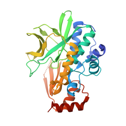
Structure of Mesorhizobium Loti Arylamine N-Acetyltransferase 1.

Holton, S.J.Dairou, J.Sandy, J.Rodrigues-Lima, F.Dupret, J.M.Noble, M.E.M.Sim, E.

(2005) Acta Crystallogr Sect F Struct Biol Cryst Commun 61: 14

  • DOI: https://doi.org/10.1107/S1744309104030659
  • Primary Citation Related Structures: 
    2BSZ

  • PubMed Abstract: 

    The arylamine N-acetyltransferase (NAT) enzymes have been found in a broad range of both eukaryotic and prokaryotic organisms. The NAT enzymes catalyse the transfer of an acetyl group from acetyl Co-enzyme A onto the terminal nitrogen of a range of arylamine, hydrazine and arylhydrazine compounds. Recently, several NAT structures have been reported from different prokaryotic sources including Salmonella typhimurium, Mycobacterium smegmatis and Pseudomonas aeruginosa. Bioinformatics analysis of the Mesorhizobium loti genome revealed two NAT paralogues, the first example of multiple NAT isoenzymes in a eubacterial organism. The M. loti NAT 1 enzyme was recombinantly expressed and purified for X-ray crystallographic studies. The purified enzyme was crystallized in 0.5 M Ca(OAc)2, 16% PEG 3350, 0.1 M Tris-HCl pH 8.5 using the sitting-drop vapour-diffusion method. A data set diffracting to 2.0 A was collected from a single crystal at 100 K. The crystal belongs to the orthorhombic spacegroup P2(1)2(1)2(1), with unit-cell parameters a = 53.2, b = 97.3, c = 114.3 A. The structure was refined to a final free-R factor of 24.8%. The structure reveals that despite low sequence homology, M. loti NAT1 shares the common fold as reported in previous NAT structures and exhibits the same catalytic triad of residues (Cys-His-Asp) in the active site.


  • Organizational Affiliation
    • Department of Biochemistry, Oxford University, Oxford OX1 3QU, England.

Macromolecule Content 

  • Total Structure Weight: 61.57 kDa 
  • Atom Count: 4,430 
  • Modeled Residue Count: 531 
  • Deposited Residue Count: 556 
  • Unique protein chains: 1

Macromolecules

Find similar proteins by:|  3D Structure
Entity ID: 1
MoleculeChains  Sequence LengthOrganismDetailsImage
ARYLAMINE N-ACETYLTRANSFERASE 1
A, B
278Mesorhizobium lotiMutation(s): 0 
EC: 2.3.1.5
UniProt
Find proteins for Q98D42 (Mesorhizobium japonicum (strain LMG 29417 / CECT 9101 / MAFF 303099))
Explore Q98D42 
Go to UniProtKB:  Q98D42
Entity Groups
Sequence Clusters30% Identity50% Identity70% Identity90% Identity95% Identity100% Identity
UniProt GroupQ98D42
Sequence Annotations
Expand
Reference Sequence

Experimental Data & Validation

Experimental Data

  • Method: X-RAY DIFFRACTION
  • Resolution: 2.00 Å
  • R-Value Free:  0.249 (Depositor), 0.243 (DCC) 
  • R-Value Work:  0.195 (Depositor), 0.199 (DCC) 
  • R-Value Observed: 0.198 (Depositor) 
Space Group: P 21 21 21
Unit Cell:
Length ( Å )Angle ( ˚ )
a = 53.239α = 90
b = 97.304β = 90
c = 114.346γ = 90
Software Package:
Software NamePurpose
REFMACrefinement
MOSFLMdata reduction
SCALAdata scaling

Structure Validation

View Full Validation Report


Entry History 

Deposition Data

Revision History  (Full details and data files)

  • Version 1.0: 2005-05-25
    Type: Initial release
  • Version 1.1: 2013-03-27
    Changes: Derived calculations, Non-polymer description, Other, Structure summary, Version format compliance
  • Version 1.2: 2023-12-13
    Changes: Data collection, Database references, Other, Refinement description