2BOY

Crystal structure of 3-ChloroCatechol 1,2-Dioxygenase from Rhodococcus Opacus 1CP


Experimental Data Snapshot

  • Method: X-RAY DIFFRACTION
  • Resolution: 1.90 Å
  • R-Value Free: 0.219 
  • R-Value Work: 0.176 
  • R-Value Observed: 0.178 

wwPDB Validation   3D Report Full Report


Ligand Structure Quality Assessment 


This is version 1.7 of the entry. See complete history


Literature

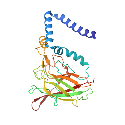
Crystal structure of 3-chlorocatechol 1,2-dioxygenase key enzyme of a new modified ortho-pathway from the Gram-positive Rhodococcus opacus 1CP grown on 2-chlorophenol.

Ferraroni, M.Kolomytseva, M.P.Solyanikova, I.P.Scozzafava, A.Golovleva, L.A.Briganti, F.

(2006) J Mol Biol 360: 788-799

  • DOI: https://doi.org/10.1016/j.jmb.2006.05.046
  • Primary Citation of Related Structures:  
    2BOY

  • PubMed Abstract: 

    The crystal structure of the 3-chlorocatechol 1,2-dioxygenase from the Gram-positive bacterium Rhodococcus opacus (erythropolis) 1CP, a Fe(III) ion-containing enzyme specialized in the aerobic biodegradation of 3-chloro- and methyl-substituted catechols, has been solved by molecular replacement techniques using the coordinates of 4-chlorocatechol 1,2-dioxygenase from the same organism (PDB code 1S9A) as a starting model and refined at 1.9 A resolution (R(free) 21.9%; R-factor 17.4%). The analysis of the structure and of the kinetic parameters for a series of different substrates, and the comparison with the corresponding data for the 4-chlorocatechol 1,2-dioxygenase isolated from the same bacterial strain, provides evidence of which active site residues are responsible for the observed differences in substrate specificity. Among the amino acid residues expected to interact with substrates, only three are altered Val53(Ala53), Tyr78(Phe78) and Ala221(Cys224) (3-chlorocatechol 1,2-dioxygenase(4-chlorocatechol 1,2-dioxygenase)), clearly identifying the substitutions influencing substrate selectivity in these enzymes. The crystallographic asymmetric unit contains eight subunits (corresponding to four dimers) that show heterogeneity in the conformation of a co-crystallized molecule bound to the catalytic non-heme iron(III) ion resembling a benzohydroxamate moiety, probably a result of the breakdown of recently discovered siderophores synthesized by Gram-positive bacteria. Several different modes of binding benzohydroxamate into the active site induce distinct conformations of the interacting protein ligands Tyr167 and Arg188, illustrating the plasticity of the active site origin of the more promiscuous substrate preferences of the present enzyme.


  • Organizational Affiliation

    Dipartimento di Chimica, Università di Firenze, Via della Lastruccia 3, I-50019 Sesto Fiorentino (FI), Italy.


Macromolecules
Find similar proteins by:  (by identity cutoff)  |  3D Structure
Entity ID: 1
MoleculeChains Sequence LengthOrganismDetailsImage
3-CHLOROCATECHOL 1,2-DIOXYGENASE
A, B, C, D, E
A, B, C, D, E, F, G, H
254Rhodococcus opacusMutation(s): 0 
EC: 1.13.11.1
UniProt
Find proteins for Q8G9L3 (Rhodococcus opacus)
Explore Q8G9L3 
Go to UniProtKB:  Q8G9L3
Entity Groups  
Sequence Clusters30% Identity50% Identity70% Identity90% Identity95% Identity100% Identity
UniProt GroupQ8G9L3
Sequence Annotations
Expand
  • Reference Sequence
Small Molecules
Ligands 4 Unique
IDChains Name / Formula / InChI Key2D Diagram3D Interactions
LPP
Query on LPP

Download Ideal Coordinates CCD File 
CA [auth D]
HA [auth E]
K [auth A]
LA [auth F]
P [auth B]
CA [auth D],
HA [auth E],
K [auth A],
LA [auth F],
P [auth B],
PA [auth G],
TA [auth H],
V [auth C]
2-(HEXADECANOYLOXY)-1-[(PHOSPHONOOXY)METHYL]ETHYL HEXADECANOATE
C35 H69 O8 P
PORPENFLTBBHSG-MGBGTMOVSA-N
BHO
Query on BHO

Download Ideal Coordinates CCD File 
BA [auth D]
GA [auth E]
J [auth A]
KA [auth F]
O [auth B]
BA [auth D],
GA [auth E],
J [auth A],
KA [auth F],
O [auth B],
OA [auth G],
SA [auth H],
U [auth C]
BENZHYDROXAMIC ACID
C7 H7 N O2
VDEUYMSGMPQMIK-UHFFFAOYSA-N
FE
Query on FE

Download Ideal Coordinates CCD File 
AA [auth D]
FA [auth E]
I [auth A]
JA [auth F]
N [auth B]
AA [auth D],
FA [auth E],
I [auth A],
JA [auth F],
N [auth B],
NA [auth G],
RA [auth H],
T [auth C]
FE (III) ION
Fe
VTLYFUHAOXGGBS-UHFFFAOYSA-N
MG
Query on MG

Download Ideal Coordinates CCD File 
DA [auth D]
EA [auth D]
IA [auth E]
L [auth A]
M [auth A]
DA [auth D],
EA [auth D],
IA [auth E],
L [auth A],
M [auth A],
MA [auth F],
Q [auth B],
QA [auth G],
R [auth B],
S [auth B],
UA [auth H],
VA [auth H],
W [auth C],
X [auth C],
Y [auth C],
Z [auth C]
MAGNESIUM ION
Mg
JLVVSXFLKOJNIY-UHFFFAOYSA-N
Experimental Data & Validation

Experimental Data

  • Method: X-RAY DIFFRACTION
  • Resolution: 1.90 Å
  • R-Value Free: 0.219 
  • R-Value Work: 0.176 
  • R-Value Observed: 0.178 
  • Space Group: P 1
Unit Cell:
Length ( Å )Angle ( ˚ )
a = 83.179α = 85.37
b = 86.61β = 66.53
c = 93.447γ = 76.94
Software Package:
Software NamePurpose
REFMACrefinement
DENZOdata reduction
SCALEPACKdata scaling
MOLREPphasing

Structure Validation

View Full Validation Report


Ligand Structure Quality Assessment 


Entry History 

Deposition Data

Revision History  (Full details and data files)

  • Version 1.0: 2006-08-24
    Type: Initial release
  • Version 1.1: 2011-05-08
    Changes: Version format compliance
  • Version 1.2: 2011-07-13
    Changes: Version format compliance
  • Version 1.3: 2018-06-06
    Changes: Data collection, Database references, Derived calculations
  • Version 1.4: 2018-12-19
    Changes: Advisory, Data collection, Derived calculations
  • Version 1.5: 2019-05-08
    Changes: Data collection, Experimental preparation
  • Version 1.6: 2019-05-29
    Changes: Data collection, Other
  • Version 1.7: 2023-12-13
    Changes: Data collection, Database references, Derived calculations, Refinement description