2BOA | pdb_00002boa

Human procarboxypeptidase A4.


Experimental Data Snapshot

  • Method: X-RAY DIFFRACTION
  • Resolution: 2.20 Å
  • R-Value Free: 
    0.293 (Depositor) 
  • R-Value Work: 
    0.220 (Depositor) 
  • R-Value Observed: 
    0.221 (Depositor) 

Starting Model: experimental
View more details

wwPDB Validation 3D Report Full Report

Validation slider image for 2BOA

This is version 2.2 of the entry. See complete history

Literature

Detailed Molecular Comparison between the Inhibition Mode of A/B-Type Carboxypeptidases in the Zymogen State and by the Endogenous Inhibitor Latexin.

Garcia-Castellanos, R.Bonet-Figueredo, R.Pallares, I.Ventura, S.Aviles, F.X.Vendrell, J.Gomis-Ruth, F.X.

(2005) Cell Mol Life Sci 62: 1996

  • DOI: https://doi.org/10.1007/s00018-005-5174-4
  • Primary Citation Related Structures: 
    2BOA

  • PubMed Abstract: 

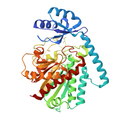
    Treatment of advanced stages of prostate carcinoma with histone-deacetylase inhibitors entails expression of human procarboxypeptidase-A4 (hPCPA4). The three-dimensional structure of hPCPA4 has been solved and shows the features of related metallocarboxypeptidase zymogens, with a preformed alpha/beta/-hydrolase active-enzyme moiety (hCPA4) and an inhibiting pro-domain (PD). The protease moiety recalls a sphere, out of which a spherical cone has been cut. This results in a funnel-like structure, at the bottom of which the active-site cleft resides. The border of this funnel is shaped by loops, which are responsible for the interaction with the PD, characterised by a large interface area and relatively few contacts. Such an inhibitory mode is evocative of the recently reported structure of the human inhibitor latexin in its complex with hCPA4. The main contacting structure of latexin is similar to the one employed for PD inhibition. In both cases, active-site blocking relies mainly on a loop provided by the central part of a beta sheet.


  • Organizational Affiliation
    • Institut de Biologia Molecular de Barcelona, C.I.D. - C.S.I.C., C/Jordi Girona 18-26, 08034, Barcelona, Spain.

Macromolecule Content 

  • Total Structure Weight: 92.37 kDa 
  • Atom Count: 6,882 
  • Modeled Residue Count: 808 
  • Deposited Residue Count: 808 
  • Unique protein chains: 1

Macromolecules

Find similar proteins by:|  3D Structure
Entity ID: 1
MoleculeChains  Sequence LengthOrganismDetailsImage
CARBOXYPEPTIDASE A4
A, B
404Homo sapiensMutation(s): 0 
EC: 3.4.17
UniProt & NIH Common Fund Data Resources
Find proteins for Q9UI42 (Homo sapiens)
Explore Q9UI42 
Go to UniProtKB:  Q9UI42
PHAROS:  Q9UI42
GTEx:  ENSG00000128510 
Entity Groups
Sequence Clusters30% Identity50% Identity70% Identity90% Identity95% Identity100% Identity
UniProt GroupQ9UI42
Glycosylation
Glycosylation Sites: 1Go to GlyGen: Q9UI42-1
Sequence Annotations
Expand
Reference Sequence

Oligosaccharides

Help  
Entity ID: 2
MoleculeChains Length2D Diagram GlycosylationD Interactions
2-acetamido-2-deoxy-beta-D-glucopyranose-(1-4)-2-acetamido-2-deoxy-beta-D-glucopyranose
C
2N-Glycosylation
Glycosylation Resources
GlyTouCan: G42666HT
GlyCosmos: G42666HT
GlyGen: G42666HT

Experimental Data & Validation

Experimental Data

  • Method: X-RAY DIFFRACTION
  • Resolution: 2.20 Å
  • R-Value Free:  0.293 (Depositor) 
  • R-Value Work:  0.220 (Depositor) 
  • R-Value Observed: 0.221 (Depositor) 
Space Group: C 1 2 1
Unit Cell:
Length ( Å )Angle ( ˚ )
a = 136.8α = 90
b = 87.2β = 117.2
c = 89.7γ = 90
Software Package:
Software NamePurpose
REFMACrefinement
MOSFLMdata reduction
SCALAdata scaling
AMoREphasing

Structure Validation

View Full Validation Report


Entry History 

Deposition Data

Revision History  (Full details and data files)

  • Version 1.0: 2005-12-13
    Type: Initial release
  • Version 1.1: 2011-05-08
    Changes: Version format compliance
  • Version 1.2: 2011-07-13
    Changes: Version format compliance
  • Version 1.3: 2017-07-12
    Changes: Derived calculations
  • Version 2.0: 2020-07-29
    Type: Remediation
    Reason: Carbohydrate remediation
    Changes: Atomic model, Data collection, Derived calculations, Structure summary
  • Version 2.1: 2023-12-13
    Changes: Data collection, Database references, Refinement description, Structure summary
  • Version 2.2: 2024-10-16
    Changes: Structure summary