2BO9 | pdb_00002bo9

Human carboxypeptidase A4 in complex with human latexin.


Experimental Data Snapshot

  • Method: X-RAY DIFFRACTION
  • Resolution: 1.60 Å
  • R-Value Free: 
    0.176 (Depositor) 
  • R-Value Work: 
    0.149 (Depositor) 
  • R-Value Observed: 
    0.150 (Depositor) 

Starting Model: other
View more details

wwPDB Validation 3D Report Full Report

Validation slider image for 2BO9

This is version 1.6 of the entry. See complete history

Literature

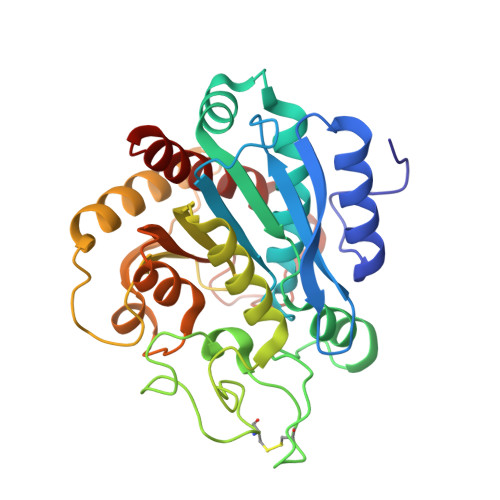
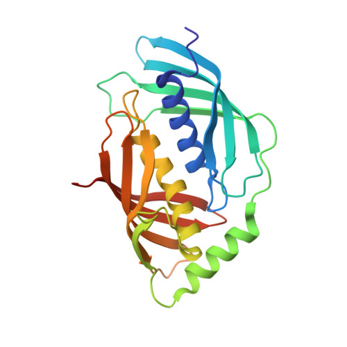
Structure of Human Carboxypeptidase A4 with its Endogenous Protein Inhibitor, Latexin.

Pallares, I.Bonet, R.Garcia-Castellanos, R.Ventura, S.Aviles, F.X.Vendrell, J.Gomis-Rueth, F.X.

(2005) Proc Natl Acad Sci U S A 102: 3978

  • DOI: https://doi.org/10.1073/pnas.0500678102
  • Primary Citation Related Structures: 
    2BO9

  • PubMed Abstract: 

    The only endogenous protein inhibitor known for metallocarboxypeptidases (MCPs) is latexin, a 25-kDa protein discovered in the rat brain. Latexin, alias endogenous carboxypeptidase inhibitor, inhibits human CPA4 (hCPA4), whose expression is induced in prostate cancer cells after treatment with histone deacetylase inhibitors. hCPA4 is a member of the A/B subfamily of MCPs and displays the characteristic alpha/beta-hydrolase fold. Human latexin consists of two topologically equivalent subdomains, reminiscent of cystatins, consisting of an alpha-helix enveloped by a curved beta-sheet. These subdomains are packed against each other through the helices and linked by a connecting segment encompassing a third alpha-helix. The enzyme is bound at the interface of these subdomains. The complex occludes a large contact surface but makes rather few contacts, despite a nanomolar inhibition constant. This low specificity explains the flexibility of latexin in inhibiting all vertebrate A/B MCPs tested, even across species barriers. In contrast, modeling studies reveal why the N/E subfamily of MCPs and invertebrate A/B MCPs are not inhibited. Major differences in the loop segments shaping the border of the funnel-like access to the protease active site impede complex formation with latexin. Several sequences ascribable to diverse tissues and organs have been identified in vertebrate genomes as being highly similar to latexin. They are proposed to constitute the latexin family of potential inhibitors. Because they are ubiquitous, latexins could represent for vertebrate A/B MCPs the counterparts of tissue inhibitors of metalloproteases for matrix metalloproteinases.


  • Organizational Affiliation
    • Institut de Biologia Molecular de Barcelona, Centro de Investigación y Desarrollo-Consejo Superior de Investigaciones Científicas, C/Jordi Girona, 18-26, E-08034 Barcelona, Spain.

Macromolecule Content 

  • Total Structure Weight: 123.01 kDa 
  • Atom Count: 9,751 
  • Modeled Residue Count: 1,048 
  • Deposited Residue Count: 1,060 
  • Unique protein chains: 2

Macromolecules

Find similar proteins by:|  3D Structure
Entity ID: 1
MoleculeChains  Sequence LengthOrganismDetailsImage
CARBOXYPEPTIDASE A4
A, C
308Homo sapiensMutation(s): 0 
EC: 3.4.17
UniProt & NIH Common Fund Data Resources
Find proteins for Q9UI42 (Homo sapiens)
Explore Q9UI42 
Go to UniProtKB:  Q9UI42
PHAROS:  Q9UI42
GTEx:  ENSG00000128510 
Entity Groups
Sequence Clusters30% Identity50% Identity70% Identity90% Identity95% Identity100% Identity
UniProt GroupQ9UI42
Glycosylation
Glycosylation Sites: 1Go to GlyGen: Q9UI42-1
Sequence Annotations
Expand
Reference Sequence
Find similar proteins by:|  3D Structure
Entity ID: 2
MoleculeChains  Sequence LengthOrganismDetailsImage
HUMAN LATEXIN
B, D
222Homo sapiensMutation(s): 0 
UniProt & NIH Common Fund Data Resources
Find proteins for Q9BS40 (Homo sapiens)
Explore Q9BS40 
Go to UniProtKB:  Q9BS40
PHAROS:  Q9BS40
GTEx:  ENSG00000079257 
Entity Groups
Sequence Clusters30% Identity50% Identity70% Identity90% Identity95% Identity100% Identity
UniProt GroupQ9BS40
Sequence Annotations
Expand
Reference Sequence

Small Molecules

Ligands 5 Unique
IDChains Name / Formula / InChI Key2D Diagram3D Interactions
NAG

Query on NAG


Download:Ideal Coordinates CCD File
E [auth A],
P [auth C]
2-acetamido-2-deoxy-beta-D-glucopyranose
C8 H15 N O6
OVRNDRQMDRJTHS-FMDGEEDCSA-N
MPD

Query on MPD


Download:Ideal Coordinates CCD File
H [auth A]
I [auth A]
J [auth A]
K [auth B]
L [auth B]
H [auth A],
I [auth A],
J [auth A],
K [auth B],
L [auth B],
M [auth B],
N [auth B],
R [auth C],
S [auth D],
T [auth D],
U [auth D],
V [auth D]
(4S)-2-METHYL-2,4-PENTANEDIOL
C6 H14 O2
SVTBMSDMJJWYQN-YFKPBYRVSA-N
VAL

Query on VAL


Download:Ideal Coordinates CCD File
G [auth A],
O [auth C]
VALINE
C5 H11 N O2
KZSNJWFQEVHDMF-BYPYZUCNSA-N
ZN

Query on ZN


Download:Ideal Coordinates CCD File
F [auth A],
Q [auth C]
ZINC ION
Zn
PTFCDOFLOPIGGS-UHFFFAOYSA-N
ACN

Query on ACN


Download:Ideal Coordinates CCD File
W [auth D]ACETONE
C3 H6 O
CSCPPACGZOOCGX-UHFFFAOYSA-N

Experimental Data & Validation

Experimental Data

  • Method: X-RAY DIFFRACTION
  • Resolution: 1.60 Å
  • R-Value Free:  0.176 (Depositor) 
  • R-Value Work:  0.149 (Depositor) 
  • R-Value Observed: 0.150 (Depositor) 
Space Group: P 1 21 1
Unit Cell:
Length ( Å )Angle ( ˚ )
a = 78.922α = 90
b = 96.651β = 99.6
c = 92.358γ = 90
Software Package:
Software NamePurpose
DMmodel building
SCALAdata scaling
AMoREphasing
MLPHAREphasing
SIGMAAphasing
DMphasing
REFMACrefinement

Structure Validation

View Full Validation Report


Entry History 

Deposition Data

Revision History  (Full details and data files)

  • Version 1.0: 2005-04-15
    Type: Initial release
  • Version 1.1: 2011-05-08
    Changes: Version format compliance
  • Version 1.2: 2011-07-13
    Changes: Version format compliance
  • Version 1.3: 2017-07-12
    Changes: Derived calculations
  • Version 1.4: 2020-07-29
    Type: Remediation
    Reason: Carbohydrate remediation
    Changes: Data collection, Derived calculations, Structure summary
  • Version 1.5: 2024-05-01
    Changes: Data collection, Database references, Refinement description, Structure summary
  • Version 1.6: 2024-11-13
    Changes: Structure summary