The Crystal Structure of Pyrococcus Furiosus Ump Kinase Provides Insight Into Catalysis and Regulation in Microbial Pyrimidine Nucleotide Biosynthesis.
Marco-Marin, C., Gil-Ortiz, F., Rubio, V.(2005) J Mol Biology 352: 438
- PubMed: 16095620 Search on PubMed
- DOI: https://doi.org/10.1016/j.jmb.2005.07.045
- Primary Citation Related Structures:
2BMU, 2BRI, 2BRX - PubMed Abstract:
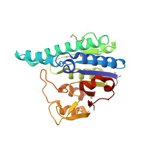
UMP kinase (UMPK), the enzyme responsible for microbial UMP phosphorylation, plays a key role in pyrimidine nucleotide biosynthesis, regulating this process via feed-back control and via gene repression of carbamoyl phosphate synthetase (the first enzyme of the pyrimidine biosynthesis pathway). We present crystal structures of Pyrococcus furiosus UMPK, free or complexed with AMPPNP or AMPPNP and UMP, at 2.4 A, 3 A and 2.55 A resolution, respectively, providing a true snapshot of the catalytically competent bisubstrate complex. The structure proves that UMPK does not resemble other nucleoside monophosphate kinases, including the UMP/CMP kinase found in animals, and thus UMPK may be a potential antimicrobial target. This enzyme has a homohexameric architecture centred around a hollow nucleus, and is organized as a trimer of dimers. The UMPK polypeptide exhibits the amino acid kinase family (AAKF) fold that has been reported in carbamate kinase and acetylglutamate kinase. Comparison with acetylglutamate kinase reveals that the substrates bind within each subunit at equivalent, adequately adapted sites. The UMPK structure contains two bound Mg ions, of which one helps stabilize the transition state, thus having the same catalytic role as one lysine residue found in acetylglutamate kinase, which is missing from P.furiosus UMPK. Relative to carbamate kinase and acetylglutamate kinase, UMPK presents a radically different dimer architecture, lacking the characteristic 16-stranded beta-sheet backbone that was considered a signature of AAKF enzymes. Its hexameric architecture, also a novel trait, results from equatorial contacts between the A and B subunits of adjacent dimers combined with polar contacts between A or B subunits, and may be required for the UMPK regulatory functions, such as gene regulation, proposed here to be mediated by hexamer-hexamer interactions with the DNA-binding protein PepA.
- Instituto de Biomedicina de Valencia (IBV-CSIC), Jaume Roig 11,Valencia 46010, Spain.
Organizational Affiliation: