2BMM | pdb_00002bmm

X-ray structure of a novel thermostable hemoglobin from the actinobacterium Thermobifida fusca


Experimental Data Snapshot

  • Method: X-RAY DIFFRACTION
  • Resolution: 2.48 Å
  • R-Value Free: 
    0.315 (Depositor), 0.205 (DCC) 
  • R-Value Work: 
    0.218 (Depositor), 0.225 (DCC) 
  • R-Value Observed: 
    0.223 (Depositor) 

Starting Model: experimental
View more details

wwPDB Validation 3D Report Full Report

Validation slider image for 2BMM

Ligand Structure Quality Assessment 


This is version 1.3 of the entry. See complete history

Literature

A Novel Thermostable Hemoglobin from the Actinobacterium Thermobifida Fusca.

Bonamore, A.Ilari, A.Giangiacomo, L.Bellelli, A.Morea, V.Boffi, A.

(2005) FEBS J 272: 4189

  • DOI: https://doi.org/10.1111/j.1742-4658.2005.04831.x
  • Primary Citation Related Structures: 
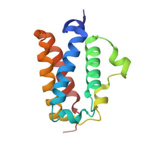
    2BMM

  • PubMed Abstract: 

    The gene coding for a hemoglobin-like protein (Tf-trHb) has been identified in the thermophilic actinobacterium Thermobifida fusca and cloned in Escherichia coli for overexpression. The crystal structure of the ferric, acetate-bound derivative, was obtained at 2.48 A resolution. The three-dimensional structure of Tf-trHb is similar to structures reported for the truncated hemoglobins from Mycobacterium tuberculosis and Bacillus subtilis in its central domain. The complete lack of diffraction patterns relative to the N- and C-terminal segments indicates that these are unstructured polypeptides chains, consistent with their facile cleavage in solution. The absence of internal cavities and the presence of two water molecules between the bound acetate ion and the protein surface suggest that the mode of ligand entry is similar to that of typical hemoglobins. The protein is characterized by higher thermostability than the similar mesophilic truncated hemoglobin from B. subtilis, as demonstrated by far-UV CD melting experiments on the cyano-met derivatives. The ligand-binding properties of Tf-trHb, analyzed in stopped flow experiments, demonstrate that Tf-trHb is capable of efficient O2 binding and release between 55 and 60 degrees C, the optimal growth temperature for Thermobifida fusca.


  • Organizational Affiliation
    • Department of Biochemical Sciences, University of Rome La Sapienza, Italy.

Macromolecule Content 

  • Total Structure Weight: 15.32 kDa 
  • Atom Count: 1,142 
  • Modeled Residue Count: 123 
  • Deposited Residue Count: 123 
  • Unique protein chains: 1

Macromolecules

Find similar proteins by:|  3D Structure
Entity ID: 1
MoleculeChains  Sequence LengthOrganismDetailsImage
THERMOSTABLE HEMOGLOBIN FROM THERMOBIFIDA FUSCA123Thermobifida fuscaMutation(s): 0 

Small Molecules

Experimental Data & Validation

Experimental Data

  • Method: X-RAY DIFFRACTION
  • Resolution: 2.48 Å
  • R-Value Free:  0.315 (Depositor), 0.205 (DCC) 
  • R-Value Work:  0.218 (Depositor), 0.225 (DCC) 
  • R-Value Observed: 0.223 (Depositor) 
Space Group: P 64 2 2
Unit Cell:
Length ( Å )Angle ( ˚ )
a = 84.617α = 90
b = 84.617β = 90
c = 77.921γ = 120
Software Package:
Software NamePurpose
REFMACrefinement
DENZOdata reduction
SCALEPACKdata scaling
MOLREPphasing

Structure Validation

View Full Validation Report


Ligand Structure Quality Assessment 


Entry History 

Deposition Data

Revision History  (Full details and data files)

  • Version 1.0: 2005-07-20
    Type: Initial release
  • Version 1.1: 2011-05-08
    Changes: Version format compliance
  • Version 1.2: 2011-07-13
    Changes: Version format compliance
  • Version 1.3: 2023-12-13
    Changes: Data collection, Database references, Other, Refinement description