2BL1 | pdb_00002bl1

Crystal structure of a putative phosphinothricin Acetyltransferase (PA4866) from Pseudomonas aeruginosa PAC1


Experimental Data Snapshot

  • Method: X-RAY DIFFRACTION
  • Resolution: 2.00 Å
  • R-Value Free: 
    0.229 (Depositor), 0.195 (DCC) 
  • R-Value Work: 
    0.197 (Depositor), 0.195 (DCC) 
  • R-Value Observed: 
    0.197 (Depositor) 

wwPDB Validation 3D Report Full Report

Validation slider image for 2BL1

This is version 1.5 of the entry. See complete history

Literature

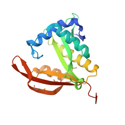
Crystal Structure of a Putative Phosphinothricin Acetyltransferase (Pa4866) from Pseudomonas Aeruginosa Pac1

Davies, A.M.Tata, R.Agha, R.Sutton, B.J.Brown, P.R.

(2005) Proteins 61: 677

Macromolecule Content 

  • Total Structure Weight: 19.47 kDa 
  • Atom Count: 1,582 
  • Modeled Residue Count: 170 
  • Deposited Residue Count: 172 
  • Unique protein chains: 1

Macromolecules

Find similar proteins by:|  3D Structure
Entity ID: 1
MoleculeChains  Sequence LengthOrganismDetailsImage
PUTATIVE PHOSPHINOTHRICIN N-ACETYLTRANSFERASE PA4866172Pseudomonas aeruginosaMutation(s): 1 
EC: 2.3.1
UniProt
Find proteins for Q9HUU7 (Pseudomonas aeruginosa (strain ATCC 15692 / DSM 22644 / CIP 104116 / JCM 14847 / LMG 12228 / 1C / PRS 101 / PAO1))
Explore Q9HUU7 
Go to UniProtKB:  Q9HUU7
Entity Groups
Sequence Clusters30% Identity50% Identity70% Identity90% Identity95% Identity100% Identity
UniProt GroupQ9HUU7
Sequence Annotations
Expand
Reference Sequence

Small Molecules

Ligands 3 Unique
IDChains Name / Formula / InChI Key2D Diagram3D Interactions
SO4

Query on SO4


Download:Ideal Coordinates CCD File
H [auth A]SULFATE ION
O4 S
QAOWNCQODCNURD-UHFFFAOYSA-L
GOL

Query on GOL


Download:Ideal Coordinates CCD File
D [auth A],
E [auth A],
F [auth A],
G [auth A]
GLYCEROL
C3 H8 O3
PEDCQBHIVMGVHV-UHFFFAOYSA-N
AZI

Query on AZI


Download:Ideal Coordinates CCD File
B [auth A],
C [auth A]
AZIDE ION
N3
IVRMZWNICZWHMI-UHFFFAOYSA-N
Modified Residues  1 Unique
IDChains TypeFormula2D DiagramParent
MSE
Query on MSE
A
L-PEPTIDE LINKINGC5 H11 N O2 SeMET

Experimental Data & Validation

Experimental Data

  • Method: X-RAY DIFFRACTION
  • Resolution: 2.00 Å
  • R-Value Free:  0.229 (Depositor), 0.195 (DCC) 
  • R-Value Work:  0.197 (Depositor), 0.195 (DCC) 
  • R-Value Observed: 0.197 (Depositor) 
Space Group: P 43 21 2
Unit Cell:
Length ( Å )Angle ( ˚ )
a = 79.99α = 90
b = 79.99β = 90
c = 61.73γ = 90
Software Package:
Software NamePurpose
CNSrefinement
DENZOdata reduction
SCALAdata scaling
SOLVEphasing

Structure Validation

View Full Validation Report


Entry History 

Deposition Data

Revision History  (Full details and data files)

  • Version 1.0: 2005-09-21
    Type: Initial release
  • Version 1.1: 2011-05-08
    Changes: Version format compliance
  • Version 1.2: 2011-07-13
    Changes: Version format compliance
  • Version 1.3: 2019-05-08
    Changes: Data collection, Derived calculations, Experimental preparation, Other
  • Version 1.4: 2019-06-12
    Changes: Data collection, Database references
  • Version 1.5: 2024-11-13
    Changes: Data collection, Database references, Other, Structure summary