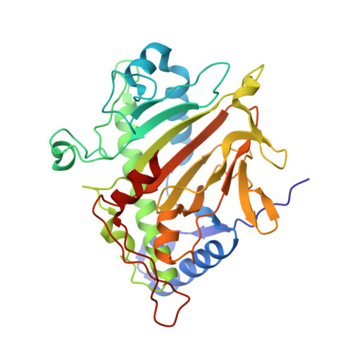
2BJS

Isopenicillin N synthase C-terminal truncation mutant


Experimental Data Snapshot

  • Method: X-RAY DIFFRACTION
  • Resolution: 1.30 Å
  • R-Value Free: 0.164 
  • R-Value Work: 0.139 
  • R-Value Observed: 0.140 

Starting Model: experimental
View more details

wwPDB Validation   3D Report Full Report


This is version 1.5 of the entry. See complete history


Literature

Terminally Truncated Isopenicillin N Synthase Generates a Dithioester Product: Evidence for a Thioaldehyde Intermediate during Catalysis and a New Mode of Reaction for Non-Heme Iron Oxidases.

McNeill, L.A.Brown, T.J.N.Sami, M.Clifton, I.J.Burzlaff, N.I.Claridge, T.D.W.Adlington, R.M.Baldwin, J.E.Rutledge, P.J.Schofield, C.J.

(2017) Chemistry 23: 12815-12824

  • DOI: https://doi.org/10.1002/chem.201701592
  • Primary Citation of Related Structures:  
    2BJS

  • PubMed Abstract: 

    Isopenicillin N synthase (IPNS) catalyses the four-electron oxidation of a tripeptide, l-δ-(α-aminoadipoyl)-l-cysteinyl-d-valine (ACV), to give isopenicillin N (IPN), the first-formed β-lactam in penicillin and cephalosporin biosynthesis. IPNS catalysis is dependent upon an iron(II) cofactor and oxygen as a co-substrate. In the absence of substrate, the carbonyl oxygen of the side-chain amide of the penultimate residue, Gln330, co-ordinates to the active-site metal iron. Substrate binding ablates the interaction between Gln330 and the metal, triggering rearrangement of seven C-terminal residues, which move to take up a conformation that extends the final α-helix and encloses ACV in the active site. Mutagenesis studies are reported, which probe the role of the C-terminal and other aspects of the substrate binding pocket in IPNS. The hydrophobic nature of amino acid side-chains around the ACV binding pocket is important in catalysis. Deletion of seven C-terminal residues exposes the active site and leads to formation of a new type of thiol oxidation product. The isolated product is shown by LC-MS and NMR analyses to be the ene-thiol tautomer of a dithioester, made up from two molecules of ACV linked between the thiol sulfur of one tripeptide and the oxidised cysteinyl β-carbon of the other. A mechanism for its formation is proposed, supported by an X-ray crystal structure, which shows the substrate ACV bound at the active site, its cysteinyl β-carbon exposed to attack by a second molecule of substrate, adjacent. Formation of this product constitutes a new mode of reaction for IPNS and non-heme iron oxidases in general.


  • Organizational Affiliation

    Oxford Centre for Molecular Sciences and the Department of Chemistry, Chemistry Research Laboratory, Mansfield Road, Oxford, OX1 3TA, UK.


Macromolecules
Find similar proteins by:  (by identity cutoff)  |  3D Structure
Entity ID: 1
MoleculeChains Sequence LengthOrganismDetailsImage
ISOPENICILLIN N SYNTHETASE325Aspergillus nidulans FGSC A4Mutation(s): 0 
EC: 1.21.3.1
UniProt
Find proteins for P05326 (Emericella nidulans (strain FGSC A4 / ATCC 38163 / CBS 112.46 / NRRL 194 / M139))
Explore P05326 
Go to UniProtKB:  P05326
Entity Groups  
Sequence Clusters30% Identity50% Identity70% Identity90% Identity95% Identity100% Identity
UniProt GroupP05326
Sequence Annotations
Expand
  • Reference Sequence
Small Molecules
Ligands 4 Unique
IDChains Name / Formula / InChI Key2D Diagram3D Interactions
ACV
Query on ACV

Download Ideal Coordinates CCD File 
C [auth A],
D [auth A]
L-D-(A-AMINOADIPOYL)-L-CYSTEINYL-D-VALINE
C14 H25 N3 O6 S
BYEIJZFKOAXBBV-ATZCPNFKSA-N
SO4
Query on SO4

Download Ideal Coordinates CCD File 
F [auth A]SULFATE ION
O4 S
QAOWNCQODCNURD-UHFFFAOYSA-L
FE
Query on FE

Download Ideal Coordinates CCD File 
B [auth A]FE (III) ION
Fe
VTLYFUHAOXGGBS-UHFFFAOYSA-N
MEE
Query on MEE

Download Ideal Coordinates CCD File 
E [auth A]METHANETHIOL
C H4 S
LSDPWZHWYPCBBB-UHFFFAOYSA-N
Experimental Data & Validation

Experimental Data

  • Method: X-RAY DIFFRACTION
  • Resolution: 1.30 Å
  • R-Value Free: 0.164 
  • R-Value Work: 0.139 
  • R-Value Observed: 0.140 
  • Space Group: P 21 21 21
Unit Cell:
Length ( Å )Angle ( ˚ )
a = 47.149α = 90
b = 71.126β = 90
c = 100.991γ = 90
Software Package:
Software NamePurpose
REFMACrefinement
MOSFLMdata reduction
SCALAdata scaling

Structure Validation

View Full Validation Report


Entry History 

Deposition Data

Revision History  (Full details and data files)

  • Version 1.0: 2006-03-09
    Type: Initial release
  • Version 1.1: 2011-07-13
    Changes: Advisory, Version format compliance
  • Version 1.2: 2017-12-06
    Changes: Database references
  • Version 1.3: 2018-02-21
    Changes: Structure summary
  • Version 1.4: 2023-12-13
    Changes: Data collection, Database references, Derived calculations, Other, Refinement description
  • Version 1.5: 2024-05-08
    Changes: Source and taxonomy