2BHW | pdb_00002bhw

PEA LIGHT-HARVESTING COMPLEX II AT 2.5 ANGSTROM RESOLUTION


Experimental Data Snapshot

  • Method: X-RAY DIFFRACTION
  • Resolution: 2.50 Å
  • R-Value Free: 
    0.241 (Depositor), 0.250 (DCC) 
  • R-Value Work: 
    0.220 (Depositor), 0.232 (DCC) 
  • R-Value Observed: 
    0.220 (Depositor) 

Starting Model: other
View more details

wwPDB Validation 3D Report Full Report

Validation slider image for 2BHW

Ligand Structure Quality Assessment 


This is version 2.0 of the entry. See complete history

Literature

Mechanisms of Photoprotection and Nonphotochemical Quenching in Pea Light-Harvesting Complex at 2.5 A Resolution.

Standfuss, J.Terwisscha Van Scheltinga, A.C.Lamborghini, M.Kuehlbrandt, W.

(2005) EMBO J 24: 919

  • DOI: https://doi.org/10.1038/sj.emboj.7600585
  • Primary Citation Related Structures: 
    2BHW

  • PubMed Abstract: 

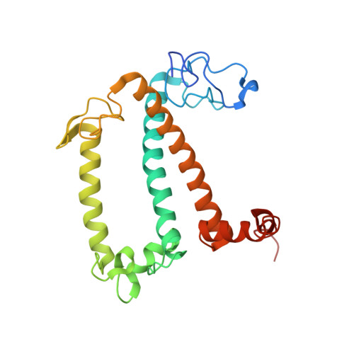
    The plant light-harvesting complex of photosystem II (LHC-II) collects and transmits solar energy for photosynthesis in chloroplast membranes and has essential roles in regulation of photosynthesis and in photoprotection. The 2.5 A structure of pea LHC-II determined by X-ray crystallography of stacked two-dimensional crystals shows how membranes interact to form chloroplast grana, and reveals the mutual arrangement of 42 chlorophylls a and b, 12 carotenoids and six lipids in the LHC-II trimer. Spectral assignment of individual chlorophylls indicates the flow of energy in the complex and the mechanism of photoprotection in two close chlorophyll a-lutein pairs. We propose a simple mechanism for the xanthophyll-related, slow component of nonphotochemical quenching in LHC-II, by which excess energy is transferred to a zeaxanthin replacing violaxanthin in its binding site, and dissipated as heat. Our structure shows the complex in a quenched state, which may be relevant for the rapid, pH-induced component of nonphotochemical quenching.


  • Organizational Affiliation
    • Max Planck Institute of Biophysics, Department of Structural Biology, Frankfurt am Main, Germany.

Macromolecule Content 

  • Total Structure Weight: 124.67 kDa 
  • Atom Count: 8,373 
  • Modeled Residue Count: 669 
  • Deposited Residue Count: 696 
  • Unique protein chains: 1

Macromolecules

Find similar proteins by:|  3D Structure
Entity ID: 1
MoleculeChains  Sequence LengthOrganismDetailsImage
CHLOROPHYLL A-B BINDING PROTEIN AB80
A, B, C
232Lathyrus oleraceusMutation(s): 0 
Membrane Entity: Yes 
UniProt
Find proteins for P07371 (Pisum sativum)
Explore P07371 
Go to UniProtKB:  P07371
Entity Groups
Sequence Clusters30% Identity50% Identity70% Identity90% Identity95% Identity100% Identity
UniProt GroupP07371
Sequence Annotations
Expand
Reference Sequence

Small Molecules

Ligands 7 Unique
IDChains Name / Formula / InChI Key2D Diagram3D Interactions
DGD

Query on DGD


Download:Ideal Coordinates CCD File
KB [auth C],
QA [auth B],
W [auth A]
DIGALACTOSYL DIACYL GLYCEROL (DGDG)
C51 H96 O15
LDQFLSUQYHBXSX-HXXRYREZSA-N
CHL

Query on CHL


Download:Ideal Coordinates CCD File
DB [auth C]
EB [auth C]
FB [auth C]
GB [auth C]
HB [auth C]
DB [auth C],
EB [auth C],
FB [auth C],
GB [auth C],
HB [auth C],
IB [auth C],
JA [auth B],
KA [auth B],
LA [auth B],
MA [auth B],
NA [auth B],
OA [auth B],
P [auth A],
Q [auth A],
R [auth A],
S [auth A],
T [auth A],
U [auth A]
CHLOROPHYLL B
C55 H70 Mg N4 O6
MWVCRINOIIOUAU-UYSPMESUSA-M
CLA

Query on CLA


Download:Ideal Coordinates CCD File
AB [auth C]
BA [auth B]
BB [auth C]
CA [auth B]
CB [auth C]
AB [auth C],
BA [auth B],
BB [auth C],
CA [auth B],
CB [auth C],
DA [auth B],
EA [auth B],
FA [auth B],
GA [auth B],
H [auth A],
HA [auth B],
I [auth A],
IA [auth B],
J [auth A],
K [auth A],
L [auth A],
M [auth A],
N [auth A],
O [auth A],
VA [auth C],
WA [auth C],
XA [auth C],
YA [auth C],
ZA [auth C]
CHLOROPHYLL A
C55 H72 Mg N4 O5
ATNHDLDRLWWWCB-AENOIHSZSA-M
LHG

Query on LHG


Download:Ideal Coordinates CCD File
JB [auth C],
PA [auth B],
V [auth A]
1,2-DIPALMITOYL-PHOSPHATIDYL-GLYCEROLE
C38 H75 O10 P
BIABMEZBCHDPBV-MPQUPPDSSA-N
NEX

Query on NEX


Download:Ideal Coordinates CCD File
F [auth A],
TA [auth C],
Z [auth B]
(1R,3R)-6-{(3E,5E,7E,9E,11E,13E,15E,17E)-18-[(1S,4R,6R)-4-HYDROXY-2,2,6-TRIMETHYL-7-OXABICYCLO[4.1.0]HEPT-1-YL]-3,7,12,16-TETRAMETHYLOCTADECA-1,3,5,7,9,11,13,15,17-NONAENYLIDENE}-1,5,5-TRIMETHYLCYCLOHEXANE-1,3-DIOL
C40 H56 O4
PGYAYSRVSAJXTE-OQASCVKESA-N
XAT

Query on XAT


Download:Ideal Coordinates CCD File
AA [auth B],
G [auth A],
UA [auth C]
(3S,5R,6S,3'S,5'R,6'S)-5,6,5',6'-DIEPOXY-5,6,5',6'- TETRAHYDRO-BETA,BETA-CAROTENE-3,3'-DIOL
C40 H56 O4
SZCBXWMUOPQSOX-WVJDLNGLSA-N
LUX

Query on LUX


Download:Ideal Coordinates CCD File
D [auth A]
E [auth A]
RA [auth C]
SA [auth C]
X [auth B]
D [auth A],
E [auth A],
RA [auth C],
SA [auth C],
X [auth B],
Y [auth B]
(3R,3'R,6'S,9R,9'R,13R,13'S)-4',5'-DIDEHYDRO-5',6',7',8',9,9',10,10',11,11',12,12',13,13',14,14',15,15'-OCTADECAHYDRO-BETA,BETA-CAROTENE-3,3'-DIOL
C40 H72 O2
OXVWOOPFXSSEKV-SQYYBNMASA-N

Experimental Data & Validation

Experimental Data

  • Method: X-RAY DIFFRACTION
  • Resolution: 2.50 Å
  • R-Value Free:  0.241 (Depositor), 0.250 (DCC) 
  • R-Value Work:  0.220 (Depositor), 0.232 (DCC) 
  • R-Value Observed: 0.220 (Depositor) 
Space Group: C 1 2 1
Unit Cell:
Length ( Å )Angle ( ˚ )
a = 211.4α = 90
b = 128β = 101.8
c = 62γ = 90
Software Package:
Software NamePurpose
TNTrefinement
XDSdata reduction
XSCALEdata scaling
MOLREPphasing

Structure Validation

View Full Validation Report


Ligand Structure Quality Assessment 


Entry History 

Deposition Data

Revision History  (Full details and data files)

  • Version 1.0: 2006-05-23
    Type: Initial release
  • Version 1.1: 2011-05-08
    Changes: Version format compliance
  • Version 1.2: 2011-07-13
    Changes: Version format compliance
  • Version 1.3: 2019-04-24
    Changes: Advisory, Data collection, Other
  • Version 2.0: 2024-05-01
    Changes: Data collection, Database references, Derived calculations, Non-polymer description, Other, Refinement description, Structure summary