Crystal Structures of Apo-Form and Binary/Ternary Complexes of Podophyllum Secoisolariciresinol Dehydrogenase, an Enzyme Involved in Formation of Health-Protecting and Plant Defense Lignans
Youn, B., Moinuddin, S.G., Davin, L.B., Lewis, N.G., Kang, C.(2005) J Biological Chem 280: 12917
- PubMed: 15653677
- DOI: https://doi.org/10.1074/jbc.M413266200
- Primary Citation of Related Structures:
2BGK, 2BGL, 2BGM - PubMed Abstract:
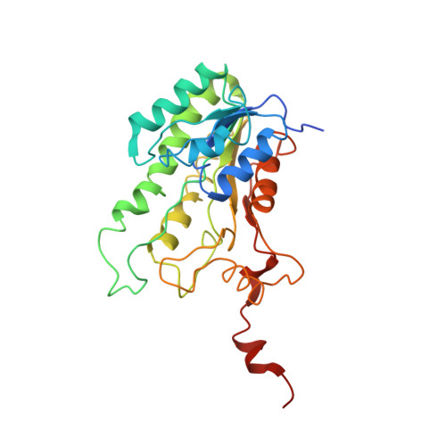
(-)-Matairesinol is a central biosynthetic intermediate to numerous 8-8'-lignans, including the antiviral agent podophyllotoxin in Podophyllum species and its semi-synthetic anticancer derivatives teniposide, etoposide, and Etopophos. It is formed by action of an enantiospecific secoisolariciresinol dehydrogenase, an NAD(H)-dependent oxidoreductase that catalyzes the conversion of (-)-secoisolariciresinol. Matairesinol is also a plant-derived precursor of the cancer-preventative "mammalian" lignan or "phytoestrogen" enterolactone, formed in the gut following ingestion of high fiber dietary foodstuffs, for example. Additionally, secoisolariciresinol dehydrogenase is involved in pathways to important plant defense molecules, such as plicatic acid in the western red cedar (Thuja plicata) heartwood. To understand the molecular and enantiospecific basis of Podophyllum secoisolariciresinol dehydrogenase, crystal structures of the apo-form and binary/ternary complexes were determined at 1.6, 2.8, and 2.0 angstrom resolution, respectively. The enzyme is a homotetramer, consisting of an alpha/beta single domain monomer containing seven parallel beta-strands flanked by eight alpha-helices on both sides. Its overall monomeric structure is similar to that of NAD(H)-dependent short-chain dehydrogenases/reductases, with a conserved Asp47 forming a hydrogen bond with both hydroxyl groups of the adenine ribose of NAD(H), and thus specificity toward NAD(H) instead of NADP(H). The highly conserved catalytic triad (Ser153, Tyr167, and Lys171) is adjacent to both NAD(+) and substrate molecules, where Tyr167 functions as a general base. Following analysis of high resolution structures of the apo-form and two complex forms, the molecular basis for both the enantio-specificity and the reaction mechanism of secoisolariciresinol dehydrogenase is discussed and compared with that of pinoresinol-lariciresinol reductase.
- School of Molecular Biosciences, Washington State University, Pullman, Washington 99164-4660, USA.
Organizational Affiliation:


