2BCV | pdb_00002bcv

DNA polymerase lambda in complex with Dttp and a DNA duplex containing an unpaired Dtmp


Experimental Data Snapshot

  • Method: X-RAY DIFFRACTION
  • Resolution: 2.00 Å
  • R-Value Free: 
    0.232 (Depositor), 0.234 (DCC) 
  • R-Value Work: 
    0.204 (Depositor), 0.208 (DCC) 
  • R-Value Observed: 
    0.204 (Depositor) 

Starting Model: experimental
View more details

wwPDB Validation 3D Report Full Report

Validation slider image for 2BCV

Ligand Structure Quality Assessment 


This is version 1.3 of the entry. See complete history

Literature

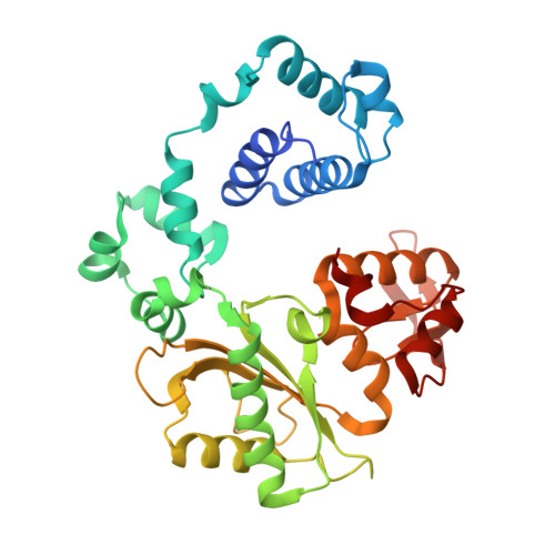
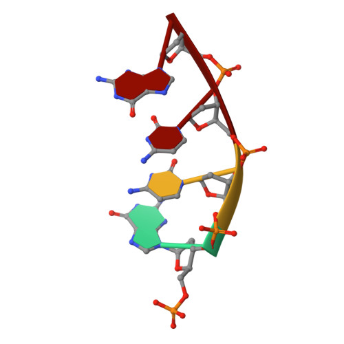
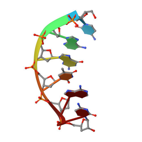
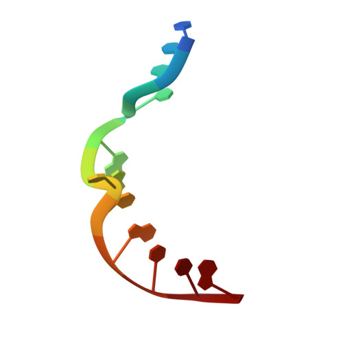
Structural analysis of strand misalignment during DNA synthesis by a human DNA polymerase

Garcia-Diaz, M.Bebenek, K.Krahn, J.M.Pedersen, L.C.Kunkel, T.A.

(2006) Cell 124: 331-342

  • DOI: https://doi.org/10.1016/j.cell.2005.10.039
  • Primary Citation Related Structures: 
    2BCQ, 2BCR, 2BCS, 2BCU, 2BCV

  • PubMed Abstract: 

    Insertions and deletions in coding sequences can alter the reading frame of genes and have profound biological consequences. In 1966, Streisinger proposed that these mutations result from strand slippage, which in repetitive sequences generates misaligned intermediates stabilized by correct base pairing that support polymerization. We report here crystal structures of human DNA polymerase lambda, which frequently generates deletion mutations, bound to such intermediates. Each contains an extrahelical template nucleotide upstream of the active site. Surprisingly, the extra nucleotide, even when combined with an adjacent mismatch, does not perturb polymerase active site geometry, which is indistinguishable from that for correctly aligned strands. These structures reveal how pol lambda can polymerize on substrates with minimal homology during repair of double-strand breaks and represent strand-slippage intermediates consistent with Streisinger's classical hypothesis. They are thus relevant to the origin of single-base deletions, a class of mutations that can confer strong biological phenotypes.


  • Organizational Affiliation
    • Laboratory of Structural Biology, National Institute of Environmental Health Sciences, NIH, DHHS, Research Triangle Park, NC 27709, USA.

Macromolecule Content 

  • Total Structure Weight: 44.72 kDa 
  • Atom Count: 3,296 
  • Modeled Residue Count: 345 
  • Deposited Residue Count: 357 
  • Unique protein chains: 1
  • Unique nucleic acid chains: 3

Macromolecules


Find similar proteins by:|  3D Structure
Entity ID: 4
MoleculeChains  Sequence LengthOrganismDetailsImage
DNA polymerase lambdaD [auth A]335Homo sapiensMutation(s): 0 
EC: 2.7.7.7 (PDB Primary Data), 4.2.99 (PDB Primary Data)
UniProt & NIH Common Fund Data Resources
Find proteins for Q9UGP5 (Homo sapiens)
Explore Q9UGP5 
Go to UniProtKB:  Q9UGP5
PHAROS:  Q9UGP5
GTEx:  ENSG00000166169 
Entity Groups
Sequence Clusters30% Identity50% Identity70% Identity90% Identity95% Identity100% Identity
UniProt GroupQ9UGP5
Sequence Annotations
Expand
Reference Sequence
Find similar nucleic acids by:  Sequence
Entity ID: 1
MoleculeChains LengthOrganismImage
5'-D(P*GP*CP*CP*G)-3'A [auth D]4N/A
Sequence Annotations
Expand
Reference Sequence
Find similar nucleic acids by:  Sequence
Entity ID: 2
MoleculeChains LengthOrganismImage
5'-D(*CP*AP*GP*TP*AP*(O2C))-3'B [auth P]6N/A
Sequence Annotations
Expand
Reference Sequence
Find similar nucleic acids by:  Sequence
Entity ID: 3
MoleculeChains LengthOrganismImage
5'-D(*CP*GP*GP*CP*AP*GP*TP*TP*AP*CP*TP*G)-3'C [auth T]12N/A
Sequence Annotations
Expand
Reference Sequence

Experimental Data & Validation

Experimental Data

  • Method: X-RAY DIFFRACTION
  • Resolution: 2.00 Å
  • R-Value Free:  0.232 (Depositor), 0.234 (DCC) 
  • R-Value Work:  0.204 (Depositor), 0.208 (DCC) 
  • R-Value Observed: 0.204 (Depositor) 
Space Group: P 21 21 21
Unit Cell:
Length ( Å )Angle ( ˚ )
a = 55.14α = 90
b = 62.988β = 90
c = 141.936γ = 90
Software Package:
Software NamePurpose
CNSrefinement
STRUCTUREdata reduction
SCALEPACKdata scaling
MOLREPphasing

Structure Validation

View Full Validation Report


Ligand Structure Quality Assessment 


Entry History 

Deposition Data

Revision History  (Full details and data files)

  • Version 1.0: 2006-03-07
    Type: Initial release
  • Version 1.1: 2008-05-01
    Changes: Version format compliance
  • Version 1.2: 2011-07-13
    Changes: Version format compliance
  • Version 1.3: 2023-08-23
    Changes: Data collection, Database references, Derived calculations, Refinement description