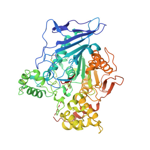
Structure of bovine pancreatic cholesterol esterase at 1.6 A: novel structural features involved in lipase activation.
Chen, J.C., Miercke, L.J., Krucinski, J., Starr, J.R., Saenz, G., Wang, X., Spilburg, C.A., Lange, L.G., Ellsworth, J.L., Stroud, R.M.(1998) Biochemistry 37: 5107-5117
- PubMed: 9548741
- DOI: https://doi.org/10.1021/bi972989g
- Primary Citation of Related Structures:
2BCE - PubMed Abstract:
The structure of pancreatic cholesterol esterase, an enzyme that hydrolyzes a wide variety of dietary lipids, mediates the absorption of cholesterol esters, and is dependent on bile salts for optimal activity, is determined to 1.6 A resolution. A full-length construct, mutated to eliminate two N-linked glycosylation sites (N187Q/N361Q), was expressed in HEK 293 cells. Enzymatic activity assays show that the purified, recombinant, mutant enzyme has activity identical to that of the native, glycosylated enzyme purified from bovine pancreas. The mutant enzyme is monomeric and exhibits improved homogeneity which aided in the growth of well-diffracting crystals. Crystals of the mutant enzyme grew in space group C2, with the following cell dimensions: a = 100.42 A, b = 54.25 A, c = 106.34 A, and beta = 104.12 degrees, with a monomer in the asymmetric unit. The high-resolution crystal structure of bovine pancreatic cholesterol esterase (Rcryst = 21.1%; Rfree = 25.0% to 1.6 A resolution) shows an alpha-beta hydrolase fold with an unusual active site environment around the catalytic triad. The hydrophobic C terminus of the protein is lodged in the active site, diverting the oxyanion hole away from the productive binding site and the catalytic Ser194. The amphipathic, helical lid found in other triglyceride lipases is truncated in the structure of cholesterol esterase and therefore is not a salient feature of activation of this lipase. These two structural features, along with the bile salt-dependent activity of the enzyme, implicate a new mode of lipase activation.
- Graduate Group in Biophysics and Department of Biochemistry and Biophysics, University of California, San Francisco, California 94143, USA.
Organizational Affiliation:


