2BBI | pdb_00002bbi

THREE-DIMENSIONAL STRUCTURE OF SOYBEAN TRYPSIN(SLASH)CHYMOTRYPSIN BOWMAN-BIRK INHIBITOR IN SOLUTION


Experimental Data Snapshot

  • Method: SOLUTION NMR
  • Conformers Submitted: 16 

wwPDB Validation 3D Report Full Report

Validation slider image for 2BBI

This is version 1.5 of the entry. See complete history

Literature

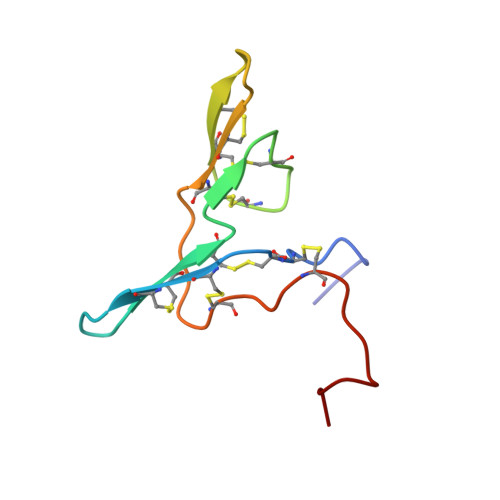
Three-dimensional structure of soybean trypsin/chymotrypsin Bowman-Birk inhibitor in solution.

Werner, M.H.Wemmer, D.E.

(1992) Biochemistry 31: 999-1010

  • DOI: https://doi.org/10.1021/bi00119a008
  • Primary Citation Related Structures: 
    1BBI, 2BBI

  • PubMed Abstract: 

    The three-dimensional structure of soybean trypsin/chymotrypsin Bowman-Birk inhibitor in solution has been determined by two-dimensional 1H nuclear magnetic resonance spectroscopy and dynamical simulated annealing using the program XPLOR. The structure was defined by 907 NOEs involving intra- and interresidue contacts which served as distance constraints for a protocol of dynamical simulated annealing. In addition, 48 phi angle constraints involving non-proline amino acids, 29 chi angle constraints, six omega angle constraints for the X-Pro peptide bond, and 35 stereoassignments for prochiral centers were incorporated during the course of the calculation. The protein is characterized by two distinct binding domains for serine protease. Each domain is comprised of a beta-hairpin (antiparallel beta-sheet and a cis-proline-containing type VIb reverse turn) with a short segment making a third strand of antiparallel beta-sheet. The structure determination and refinement are described, and the structure is compared to other structures of Bowman-Birk inhibitors as well as other families of serine protease inhibitors.


  • Organizational Affiliation
    • Chemical Biodynamics Division, Lawrence Berkeley Laboratory, University of California 94720.

Macromolecule Content 

  • Total Structure Weight: 7.88 kDa 
  • Atom Count: 536 
  • Modeled Residue Count: 71 
  • Deposited Residue Count: 71 
  • Unique protein chains: 1

Macromolecules

Find similar proteins by:|  3D Structure
Entity ID: 1
MoleculeChains  Sequence LengthOrganismDetailsImage
TRYPSIN/CHYMOTRYPSIN BOWMAN-BIRK INHIBITOR71Glycine maxMutation(s): 0 
UniProt
Find proteins for P01055 (Glycine max)
Explore P01055 
Go to UniProtKB:  P01055
Entity Groups
Sequence Clusters30% Identity50% Identity70% Identity90% Identity95% Identity100% Identity
UniProt GroupP01055
Sequence Annotations
Expand
Reference Sequence

Experimental Data & Validation

Experimental Data

  • Method: SOLUTION NMR
  • Conformers Submitted: 16 

Structure Validation

View Full Validation Report


Entry History 

Deposition Data

Revision History  (Full details and data files)

  • Version 1.0: 1993-10-31
    Type: Initial release
  • Version 1.1: 2008-03-03
    Changes: Version format compliance
  • Version 1.2: 2011-07-13
    Changes: Version format compliance
  • Version 1.3: 2013-09-25
    Changes: Derived calculations
  • Version 1.4: 2017-11-29
    Changes: Derived calculations, Other
  • Version 1.5: 2024-10-23
    Changes: Data collection, Database references, Structure summary