2BAT | pdb_00002bat

THE STRUCTURE OF THE COMPLEX BETWEEN INFLUENZA VIRUS NEURAMINIDASE AND SIALIC ACID, THE VIRAL RECEPTOR


Experimental Data Snapshot

  • Method: X-RAY DIFFRACTION
  • Resolution: 2.00 Å
  • R-Value Work: 
    0.210 (Depositor) 
  • R-Value Observed: 
    0.210 (Depositor) 

wwPDB Validation 3D Report Full Report

Validation slider image for 2BAT

This is version 2.1 of the entry. See complete history

Literature

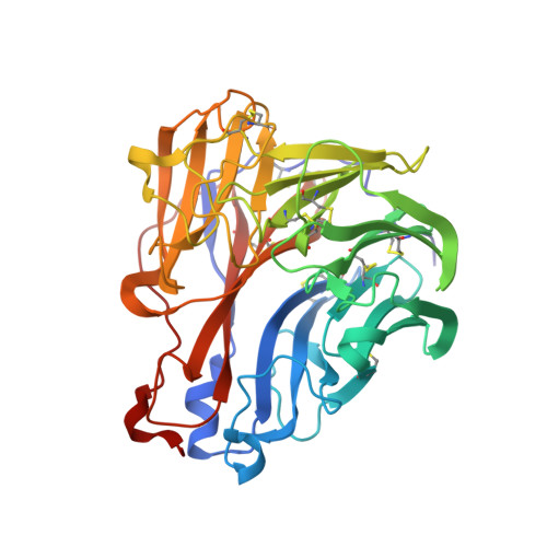
The structure of the complex between influenza virus neuraminidase and sialic acid, the viral receptor.

Varghese, J.N.McKimm-Breschkin, J.L.Caldwell, J.B.Kortt, A.A.Colman, P.M.

(1992) Proteins 14: 327-332

  • DOI: https://doi.org/10.1002/prot.340140302
  • Primary Citation Related Structures: 
    2BAT

  • PubMed Abstract: 

    Crystallographic studies of neuraminidase-sialic acid complexes indicate that sialic acid is distorted on binding the enzyme. Three arginine residues on the enzyme interact with the carboxylate group of the sugar which is observed to be equatorial to the saccharide ring as a consequence of its distorted geometry. The glycosidic oxygen is positioned within hydrogen-bonding distance of Asp-151, implicating this residue in catalysis.


  • Organizational Affiliation
    • CSIRO, Division of Biomolecular Engineering, Parkville, Victoria, Australia.

Macromolecule Content 

  • Total Structure Weight: 46.39 kDa 
  • Atom Count: 3,352 
  • Modeled Residue Count: 388 
  • Deposited Residue Count: 388 
  • Unique protein chains: 1

Macromolecules

Find similar proteins by:|  3D Structure
Entity ID: 1
MoleculeChains  Sequence LengthOrganismDetailsImage
NEURAMINIDASE N2388Influenza A virus (A/Tokyo/3/1967(H2N2))Mutation(s): 0 
EC: 3.2.1.18
UniProt
Find proteins for P06820 (Influenza A virus (strain A/Tokyo/3/1967 H2N2))
Explore P06820 
Go to UniProtKB:  P06820
Entity Groups
Sequence Clusters30% Identity50% Identity70% Identity90% Identity95% Identity100% Identity
UniProt GroupP06820
Glycosylation
Glycosylation Sites: 4
Sequence Annotations
Expand
Reference Sequence

Oligosaccharides

Help  
Entity ID: 2
MoleculeChains Length2D Diagram GlycosylationD Interactions
2-acetamido-2-deoxy-4-O-sulfo-alpha-D-galactopyranose-(1-4)-2-acetamido-2-deoxy-beta-D-glucopyranose-(1-4)-alpha-D-mannopyranose-(1-3)-beta-D-mannopyranose-(1-4)-2-acetamido-2-deoxy-beta-D-glucopyranose-(1-4)-[beta-L-fucopyranose-(1-6)]2-acetamido-2-deoxy-beta-D-glucopyranose
B
7N-Glycosylation
Glycosylation Resources
GlyTouCan: G29489WX
GlyCosmos: G29489WX
Entity ID: 3
MoleculeChains Length2D Diagram GlycosylationD Interactions
alpha-D-mannopyranose-(1-2)-alpha-D-mannopyranose-(1-3)-[alpha-D-mannopyranose-(1-6)]beta-D-mannopyranose-(1-4)-2-acetamido-2-deoxy-beta-D-glucopyranose-(1-4)-2-acetamido-2-deoxy-beta-D-glucopyranose
C
6N-Glycosylation
Glycosylation Resources
GlyTouCan: G56014GC
GlyCosmos: G56014GC
GlyGen: G56014GC

Small Molecules

Experimental Data & Validation

Experimental Data

  • Method: X-RAY DIFFRACTION
  • Resolution: 2.00 Å
  • R-Value Work:  0.210 (Depositor) 
  • R-Value Observed: 0.210 (Depositor) 
Space Group: I 4 2 2
Unit Cell:
Length ( Å )Angle ( ˚ )
a = 139.6α = 90
b = 139.6β = 90
c = 191γ = 90
Software Package:
Software NamePurpose
X-PLORmodel building
X-PLORrefinement
X-PLORphasing

Structure Validation

View Full Validation Report


Entry History 

Deposition Data

Revision History  (Full details and data files)

  • Version 1.0: 1994-01-31
    Type: Initial release
  • Version 1.1: 2008-03-24
    Changes: Version format compliance
  • Version 1.2: 2011-07-13
    Changes: Derived calculations, Version format compliance
  • Version 2.0: 2020-07-29
    Type: Remediation
    Reason: Carbohydrate remediation
    Changes: Advisory, Atomic model, Data collection, Derived calculations, Other, Structure summary
  • Version 2.1: 2024-11-20
    Changes: Advisory, Data collection, Database references, Structure summary