2AYW

Crystal Structure of the complex formed between trypsin and a designed synthetic highly potent inhibitor in the presence of benzamidine at 0.97 A resolution


Experimental Data Snapshot

  • Method: X-RAY DIFFRACTION
  • Resolution: 0.97 Å
  • R-Value Free: 0.157 
  • R-Value Work: 0.134 
  • R-Value Observed: 0.138 

Starting Model: experimental
View more details

wwPDB Validation   3D Report Full Report


Ligand Structure Quality Assessment 


This is version 1.4 of the entry. See complete history


Literature

Structure of the complex of trypsin with a highly potent synthetic inhibitor at 0.97 A resolution.

Sherawat, M.Kaur, P.Perbandt, M.Betzel, C.Slusarchyk, W.A.Bisacchi, G.S.Chang, C.Jacobson, B.L.Einspahr, H.M.Singh, T.P.

(2007) Acta Crystallogr D Biol Crystallogr 63: 500-507

  • DOI: https://doi.org/10.1107/S090744490700697X
  • Primary Citation of Related Structures:  
    2AYW

  • PubMed Abstract: 

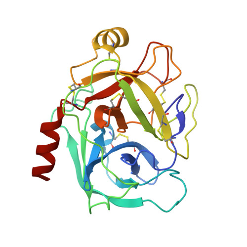
    The structure of the complex formed between bovine beta-trypsin and the highly potent synthetic inhibitor 2-{3'-[5'-methoxy-2'-(N-p-diaminomethylphenyl)amido]-1'-pyrido}-5-(N-2''-t-butylethanol)amidobenzoic acid (C(28)H(32)N(5)O(6)) has been determined at 0.97 A resolution. X-ray intensity data were collected to 0.97 A under cryocooled conditions. The structure was refined anisotropically using REFMAC5 and SHELX-97 to R(cryst) factors of 13.4 and 12.6% and R(free) factors of 15.7 and 16.3%, respectively. Several regions of the main chain and side chains that have not been previously observed were clearly defined in the present structure. H atoms are indicated as significant peaks in an |F(o) - F(c)| difference map, which accounts for an estimated 35% of all H atoms at the 2.5sigma level. The C, N and O atoms are definitively differentiated in the electron-density maps. The amido part of the inhibitor occupies the specificity pocket and the remainder fills the remaining part of the ligand-binding cleft and interacts with the enzyme through an extensive network of hydrogen bonds. The inhibitor distorts the stereochemistry of the catalytic triad, Ser195-His57-Asp102, thereby blocking the proton-relay process of the active site by preventing the formation of the crucial hydrogen bond between His57 N(delta1) and Asp102 O(delta1).


  • Organizational Affiliation

    Department of Biophysics, All India Institute of Medical Sciences, New Delhi 110029, India.


Macromolecules
Find similar proteins by:  (by identity cutoff)  |  3D Structure
Entity ID: 1
MoleculeChains Sequence LengthOrganismDetailsImage
Cationic trypsin223Bos taurusMutation(s): 0 
EC: 3.4.21.4
UniProt
Find proteins for P00760 (Bos taurus)
Explore P00760 
Go to UniProtKB:  P00760
Entity Groups  
Sequence Clusters30% Identity50% Identity70% Identity90% Identity95% Identity100% Identity
UniProt GroupP00760
Sequence Annotations
Expand
  • Reference Sequence
Small Molecules
Ligands 5 Unique
IDChains Name / Formula / InChI Key2D Diagram3D Interactions
ONO
Query on ONO

Download Ideal Coordinates CCD File 
C [auth A],
D [auth A]
2-[2-({[4-(DIAMINOMETHYL)PHENYL]AMINO}CARBONYL)-6-METHOXYPYRIDIN-3-YL]-5-{[(1-FORMYL-2,2-DIMETHYLPROPYL)AMINO]CARBONYL}BENZOIC ACID
C28 H31 N5 O6
XTFRBCPDFSHPBF-OAQYLSRUSA-N
MES
Query on MES

Download Ideal Coordinates CCD File 
F [auth A]2-(N-MORPHOLINO)-ETHANESULFONIC ACID
C6 H13 N O4 S
SXGZJKUKBWWHRA-UHFFFAOYSA-N
BEN
Query on BEN

Download Ideal Coordinates CCD File 
E [auth A]BENZAMIDINE
C7 H8 N2
PXXJHWLDUBFPOL-UHFFFAOYSA-N
GOL
Query on GOL

Download Ideal Coordinates CCD File 
G [auth A]GLYCEROL
C3 H8 O3
PEDCQBHIVMGVHV-UHFFFAOYSA-N
CA
Query on CA

Download Ideal Coordinates CCD File 
B [auth A]CALCIUM ION
Ca
BHPQYMZQTOCNFJ-UHFFFAOYSA-N
Experimental Data & Validation

Experimental Data

  • Method: X-RAY DIFFRACTION
  • Resolution: 0.97 Å
  • R-Value Free: 0.157 
  • R-Value Work: 0.134 
  • R-Value Observed: 0.138 
  • Space Group: P 32 2 1
Unit Cell:
Length ( Å )Angle ( ˚ )
a = 68.55α = 90
b = 68.55β = 90
c = 73.33γ = 120
Software Package:
Software NamePurpose
REFMACrefinement
XDSdata reduction
SCALEPACKdata scaling
AMoREphasing

Structure Validation

View Full Validation Report


Ligand Structure Quality Assessment 


Entry History 

Deposition Data

Revision History  (Full details and data files)

  • Version 1.0: 2006-01-17
    Type: Initial release
  • Version 1.1: 2008-05-01
    Changes: Version format compliance
  • Version 1.2: 2011-07-13
    Changes: Non-polymer description, Version format compliance
  • Version 1.3: 2017-10-11
    Changes: Refinement description
  • Version 1.4: 2023-10-25
    Changes: Data collection, Database references, Derived calculations, Refinement description