Kinetic, Spectroscopic, and X-ray Crystallographic Characterization of the Functional E151H Aminopeptidase from Aeromonas proteolytica.
Bzymek, K.P., Moulin, A., Swierczek, S.I., Ringe, D., Petsko, G.A., Bennett, B., Holz, R.C.(2005) Biochemistry 44: 12030-12040
- PubMed: 16142900
- DOI: https://doi.org/10.1021/bi0505823
- Primary Citation of Related Structures:
2ANP - PubMed Abstract:
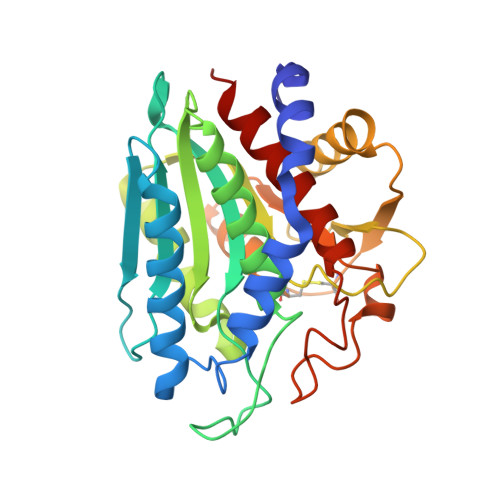
Glutamate151 (E151) has been shown to be catalytically essential for the aminopeptidase from Vibrio proteolyticus (AAP). E151 acts as the general acid/base during the catalytic mechanism of peptide hydrolysis. However, a glutamate residue is not the only residue capable of functioning as a general acid/base during catalysis for dinuclear metallohydrolases. Recent crystallographic characterization of the D-aminopeptidase from Bacillus subtilis (DppA) revealed a histidine residue that resides in an identical position to E151 in AAP. Because the active-site ligands for DppA and AAP are identical, AAP has been used as a model enzyme to understand the mechanistic role of H115 in DppA. Substitution of E151 with histidine resulted in an active AAP enzyme exhibiting a kcat value of 2.0 min(-1), which is over 2000 times slower than r AAP (4380 min(-1)). ITC experiments revealed that ZnII binds 330 and 3 times more weakly to E151H-AAP compared to r-AAP. UV-vis and EPR spectra of CoII-loaded E151H-AAP indicated that the first metal ion resides in a hexacoordinate/pentacoordinate equilibrium environment, whereas the second metal ion is six-coordinate. pH dependence of the kinetic parameters kcat and K(m) for the hydrolysis of L-leucine p-nitroanilide (L-pNA) revealed a change in an ionization constant in the enzyme-substrate complex from 5.3 in r-AAP to 6.4 in E151H-AAP, consistent with E151 in AAP being the active-site general acid/base. Proton inventory studies at pH 8.50 indicate the transfer of one proton in the rate-limiting step of the reaction. Moreover, the X-ray crystal structure of [ZnZn(E151H-AAP)] has been solved to 1.9 A resolution, and alteration of E151 to histidine does not introduce any major conformational changes to the overall protein structure or the dinuclear ZnII active site. Therefore, a histidine residue can function as the general acid/base in hydrolysis reactions of peptides and, through analogy of the role of E151 in AAP, H115 in DppA likely shuttles a proton to the leaving group of the substrate.
- Department of Chemistry and Biochemistry, Utah State University, Logan, Utah 84322-0300, USA.
Organizational Affiliation:


