2AMI | pdb_00002ami

Solution Structure Of The Calcium-loaded N-Terminal Sensor Domain Of Centrin


Experimental Data Snapshot

  • Method: SOLUTION NMR
  • Conformers Calculated: 50 
  • Conformers Submitted: 20 
  • Selection Criteria: structures with the least restraint violations,structures with the lowest energy 

wwPDB Validation 3D Report Full Report

Validation slider image for 2AMI

This is version 1.4 of the entry. See complete history

Literature

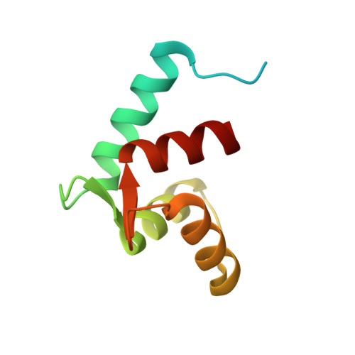
Structure of the N-terminal calcium sensor domain of centrin reveals the biochemical basis for domain-specific function.

Sheehan, J.H.Bunick, C.G.Hu, H.Fagan, P.A.Meyn, S.M.Chazin, W.J.

(2006) J Biological Chem 281: 2876-2881

  • DOI: https://doi.org/10.1074/jbc.M509886200
  • Primary Citation Related Structures: 
    2AMI

  • PubMed Abstract: 

    Centrin is an essential component of microtubule-organizing centers in organisms ranging from algae and yeast to humans. It is an EF-hand calcium-binding protein with homology to calmodulin but distinct calcium binding properties. In a previously proposed model, the C-terminal domain of centrin serves as a constitutive anchor to target proteins, and the N-terminal domain serves as the sensor of calcium signals. The three-dimensional structure of the N-terminal domain of Chlamydomonas rheinhardtii centrin has been determined in the presence of calcium by solution NMR spectroscopy. The domain is found to occupy an open conformation typical of EF-hand calcium sensors. Comparison of the N- and C-terminal domains of centrin reveals a structural and biochemical basis for the domain specificity of interactions with its cellular targets and the distinct nature of centrin relative to other EF-hand proteins. An NMR titration of the centrin N-terminal domain with a fragment of the known centrin target Sfi1 reveals binding of the peptide to a discrete site on the protein, which supports the proposal that the N-terminal domain serves as a calcium sensor in centrin.


  • Organizational Affiliation
    • Department of Biochemistry, Vanderbilt University, Nashville, Tennessee 37232-8725, USA.

Macromolecule Content 

  • Total Structure Weight: 10.82 kDa 
  • Atom Count: 601 
  • Modeled Residue Count: 76 
  • Deposited Residue Count: 96 
  • Unique protein chains: 1

Macromolecules

Find similar proteins by:|  3D Structure
Entity ID: 1
MoleculeChains  Sequence LengthOrganismDetailsImage
Caltractin96Chlamydomonas reinhardtiiMutation(s): 0 
UniProt
Find proteins for P05434 (Chlamydomonas reinhardtii)
Explore P05434 
Go to UniProtKB:  P05434
Entity Groups
Sequence Clusters30% Identity50% Identity70% Identity90% Identity95% Identity100% Identity
UniProt GroupP05434
Sequence Annotations
Expand
Reference Sequence

Experimental Data & Validation

Experimental Data

  • Method: SOLUTION NMR
  • Conformers Calculated: 50 
  • Conformers Submitted: 20 
  • Selection Criteria: structures with the least restraint violations,structures with the lowest energy 

Structure Validation

View Full Validation Report


Entry History 

Deposition Data

Revision History  (Full details and data files)

  • Version 1.0: 2005-08-23
    Type: Initial release
  • Version 1.1: 2008-04-30
    Changes: Version format compliance
  • Version 1.2: 2011-07-13
    Changes: Version format compliance
  • Version 1.3: 2014-01-22
    Changes: Database references
  • Version 1.4: 2024-05-01
    Changes: Data collection, Database references