2AID | pdb_00002aid

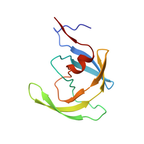
STRUCTURE OF A NON-PEPTIDE INHIBITOR COMPLEXED WITH HIV-1 PROTEASE: DEVELOPING A CYCLE OF STRUCTURE-BASED DRUG DESIGN


Experimental Data Snapshot

  • Method: X-RAY DIFFRACTION
  • Resolution: 1.90 Å
  • R-Value Work: 
    0.179 (Depositor) 
  • R-Value Observed: 
    0.179 (Depositor) 

Starting Model: experimental
View more details

wwPDB Validation 3D Report Full Report

Validation slider image for 2AID

This is version 1.6 of the entry. See complete history. 

Literature

Structure of a non-peptide inhibitor complexed with HIV-1 protease. Developing a cycle of structure-based drug design.

Rutenber, E., Fauman, E.B., Keenan, R.J., Fong, S., Furth, P.S., Ortiz de Montellano, P.R., Meng, E., Kuntz, I.D., DeCamp, D.L., Salto, R., Rose, J.R., Craik, C.S., Stroud, R.M.

(1993) J Biol Chem 268: 15343-15346

  • Primary Citation Related Structures: 
    1AID, 2AID

  • PubMed Abstract: 

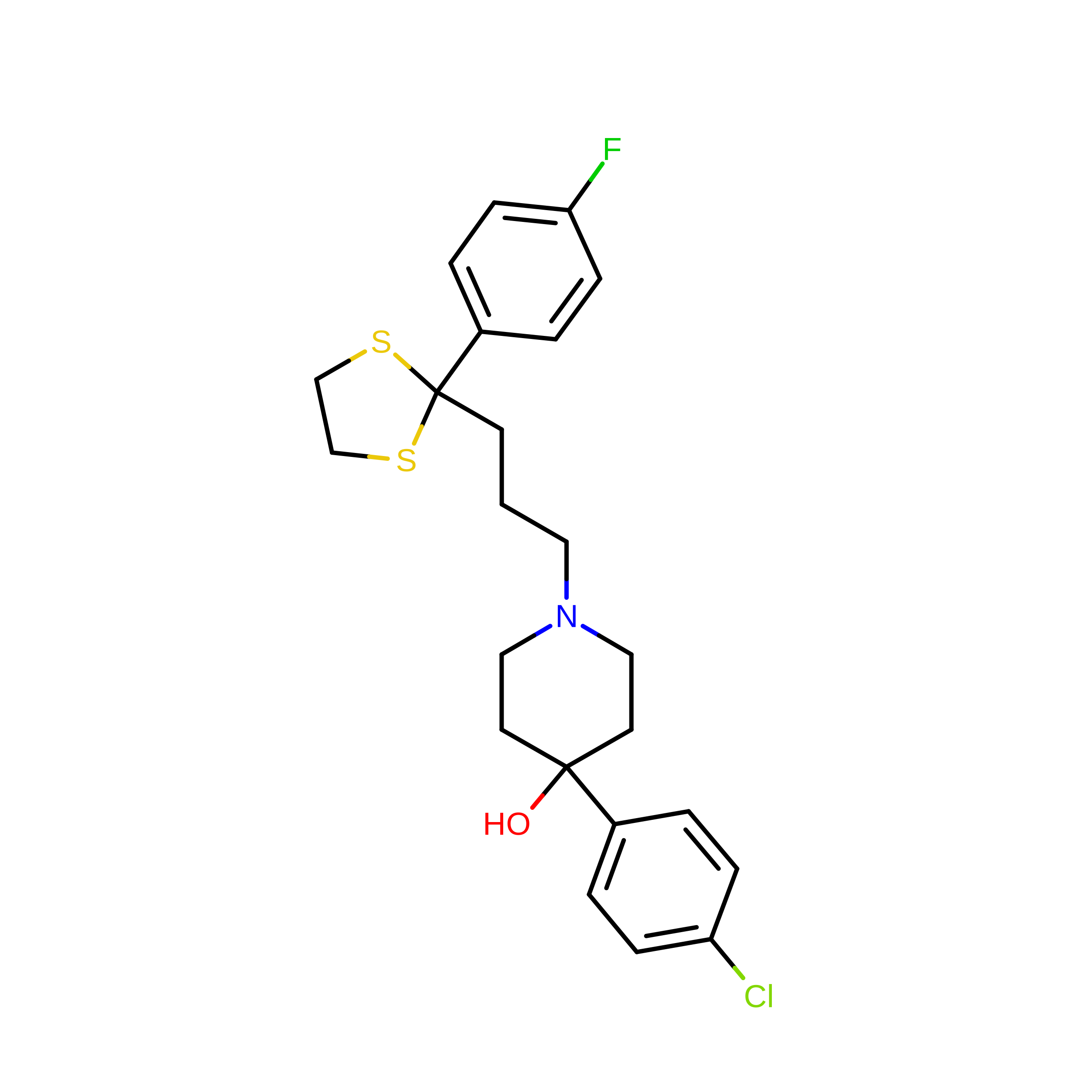
    A stable, non-peptide inhibitor of the protease from type 1 human immunodeficiency virus has been developed, and the stereochemistry of binding defined through crystallographic three-dimensional structure determination. The initial compound, haloperidol, was discovered through computational screening of the Cambridge Structural Database using a shape complementarity algorithm. The subsequent modification is a non-peptidic lateral lead, which belongs to a family of compounds with well characterized pharmacological properties. This thioketal derivative of haloperidol and a halide counterion are bound within the enzyme active site in a mode distinct from the observed for peptide-based inhibitors. A variant of the protease cocrystallized with this inhibitor shows binding in the manner predicted during the initial computer-based search. The structures provide the context for subsequent synthetic modifications of the inhibitor.


  • Organizational Affiliation: 
    • Department of Biochemistry and Biophysics, University of California, San Francisco 94143-0448.

Macromolecule Content 

  • Total Structure Weight: 22.58 kDa 
  • Atom Count: 1,682 
  • Modeled Residue Count: 198 
  • Deposited Residue Count: 198 
  • Unique protein chains: 1

Macromolecules

Find similar proteins by:|  3D Structure
Entity ID: 1
MoleculeChains  Sequence LengthOrganismDetailsImage
HUMAN IMMUNODEFICIENCY VIRUS PROTEASE
A, B
99Human immunodeficiency virus 1Mutation(s): 1 
EC: 3.4.23.16
UniProt
Find proteins for P03369 (Human immunodeficiency virus type 1 group M subtype B (isolate ARV2/SF2))
Explore P03369 
Go to UniProtKB:  P03369
Entity Groups
Sequence Clusters30% Identity50% Identity70% Identity90% Identity95% Identity100% Identity
UniProt GroupP03369
Sequence Annotations
Expand
Reference Sequence

Small Molecules

Experimental Data & Validation

Experimental Data

  • Method: X-RAY DIFFRACTION
  • Resolution: 1.90 Å
  • R-Value Work:  0.179 (Depositor) 
  • R-Value Observed: 0.179 (Depositor) 
Space Group: P 21 21 21
Unit Cell:
Length ( Å )Angle ( ˚ )
a = 51.5α = 90
b = 60.4β = 90
c = 62.2γ = 90
Software Package:
Software NamePurpose
X-PLORmodel building
X-PLORrefinement
BUDDHAdata reduction
XENGENdata scaling
X-PLORphasing

Structure Validation

View Full Validation Report


Entry History 

Deposition Data

Revision History  (Full details and data files)

  • Version 1.0: 1997-10-15
    Type: Initial release
  • Version 1.1: 2008-03-24
    Changes: Version format compliance
  • Version 1.2: 2011-07-13
    Changes: Version format compliance
  • Version 1.3: 2012-02-22
    Changes: Database references
  • Version 1.4: 2021-11-03
    Changes: Database references, Derived calculations, Other
  • Version 1.5: 2023-08-09
    Changes: Refinement description
  • Version 1.6: 2024-05-22
    Changes: Data collection