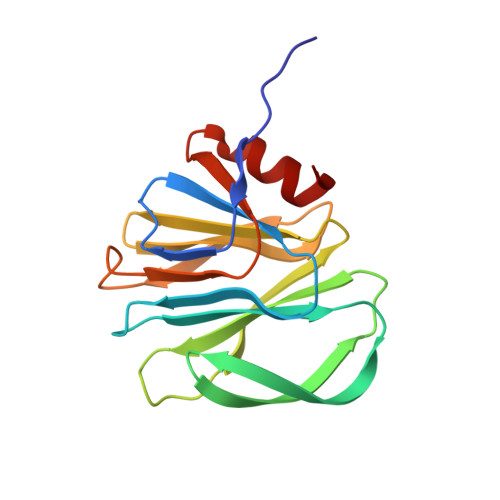
2AEN

Crystal structure of the rotavirus strain DS-1 VP8* core


Experimental Data Snapshot

  • Method: X-RAY DIFFRACTION
  • Resolution: 1.60 Å
  • R-Value Free: 0.192 
  • R-Value Work: 0.158 
  • R-Value Observed: 0.160 

Starting Model: experimental
View more details

wwPDB Validation   3D Report Full Report


This is version 1.5 of the entry. See complete history


Literature

High-resolution molecular and antigen structure of the VP8* core of a sialic acid-independent human rotavirus strain

Monnier, N.Higo-Moriguchi, K.Sun, Z.-Y.J.Prasad, B.V.V.Taniguchi, K.Dormitzer, P.R.

(2006) J Virol 80: 1513-1523

  • DOI: https://doi.org/10.1128/JVI.80.3.1513-1523.2006
  • Primary Citation of Related Structures:  
    2AEN

  • PubMed Abstract: 

    The most intensively studied rotavirus strains initially attach to cells when the "heads" of their protruding spikes bind cell surface sialic acid. Rotavirus strains that cause disease in humans do not bind this ligand. The structure of the sialic acid binding head (the VP8* core) from the simian rotavirus strain RRV has been reported, and neutralization epitopes have been mapped onto its surface. We report here a 1.6-A resolution crystal structure of the equivalent domain from the sialic acid-independent rotavirus strain DS-1, which causes gastroenteritis in humans. Although the RRV and DS-1 VP8* cores differ functionally, they share the same galectin-like fold. Differences between the RRV and DS-1 VP8* cores in the region that corresponds to the RRV sialic acid binding site make it unlikely that DS-1 VP8* binds an alternative carbohydrate ligand in this location. In the crystals, a surface cleft on each DS-1 VP8* core binds N-terminal residues from a neighboring molecule. This cleft may function as a ligand binding site during rotavirus replication. We also report an escape mutant analysis, which allows the mapping of heterotypic neutralizing epitopes recognized by human monoclonal antibodies onto the surface of the VP8* core. The distribution of escape mutations on the DS-1 VP8* core indicates that neutralizing antibodies that recognize VP8* of human rotavirus strains may bind a conformation of the spike that differs from those observed to date.


  • Organizational Affiliation

    Harvard College, Cambridge, MA 02138, USA.


Macromolecules
Find similar proteins by:  (by identity cutoff)  |  3D Structure
Entity ID: 1
MoleculeChains Sequence LengthOrganismDetailsImage
Outer capsid protein VP4, VP8* core
A, B, C, D, E
A, B, C, D, E, F, G, H
164Human rotavirus AMutation(s): 0 
Gene Names: GENOME SEGEMENT 4
UniProt
Find proteins for P11196 (Rotavirus A (strain RVA/Human/United States/DS-1/1976/G2P1B[4]))
Explore P11196 
Go to UniProtKB:  P11196
Entity Groups  
Sequence Clusters30% Identity50% Identity70% Identity90% Identity95% Identity100% Identity
UniProt GroupP11196
Sequence Annotations
Expand
  • Reference Sequence
Small Molecules
Ligands 2 Unique
IDChains Name / Formula / InChI Key2D Diagram3D Interactions
GOL
Query on GOL

Download Ideal Coordinates CCD File 
J [auth B]
K [auth B]
M [auth D]
N [auth E]
O [auth G]
J [auth B],
K [auth B],
M [auth D],
N [auth E],
O [auth G],
P [auth G]
GLYCEROL
C3 H8 O3
PEDCQBHIVMGVHV-UHFFFAOYSA-N
EOH
Query on EOH

Download Ideal Coordinates CCD File 
I [auth A],
L [auth B],
Q [auth G],
R [auth H]
ETHANOL
C2 H6 O
LFQSCWFLJHTTHZ-UHFFFAOYSA-N
Experimental Data & Validation

Experimental Data

  • Method: X-RAY DIFFRACTION
  • Resolution: 1.60 Å
  • R-Value Free: 0.192 
  • R-Value Work: 0.158 
  • R-Value Observed: 0.160 
  • Space Group: P 1
Unit Cell:
Length ( Å )Angle ( ˚ )
a = 42.421α = 90.03
b = 84.123β = 90.02
c = 90.769γ = 75.54
Software Package:
Software NamePurpose
REFMACrefinement
SCALEPACKdata scaling
CNSrefinement
HKL-2000data collection
HKL-2000data reduction
CNSphasing

Structure Validation

View Full Validation Report


Entry History 

Deposition Data

Revision History  (Full details and data files)

  • Version 1.0: 2006-02-07
    Type: Initial release
  • Version 1.1: 2008-04-30
    Changes: Version format compliance
  • Version 1.2: 2011-07-13
    Changes: Non-polymer description, Version format compliance
  • Version 1.3: 2017-12-20
    Changes: Database references
  • Version 1.4: 2019-07-24
    Changes: Data collection, Refinement description
  • Version 1.5: 2023-08-23
    Changes: Data collection, Database references, Derived calculations, Refinement description