2ADB

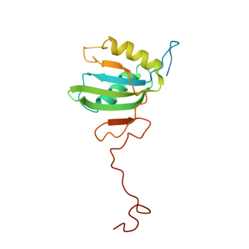
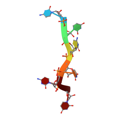
Solution structure of Polypyrimidine Tract Binding protein RBD2 complexed with CUCUCU RNA


Experimental Data Snapshot

  • Method: SOLUTION NMR
  • Conformers Calculated: 40 
  • Conformers Submitted: 20 
  • Selection Criteria: structures with the lowest energy 

wwPDB Validation   3D Report Full Report


This is version 1.4 of the entry. See complete history


Literature

Structure of PTB bound to RNA: specific binding and implications for splicing regulation

Oberstrass, F.C.Auweter, S.D.Erat, M.Hargous, Y.Henning, A.Wenter, P.Reymond, L.Amir-Ahmady, B.Pitsch, S.Black, D.L.Allain, F.H.T.

(2005) Science 309: 2054-2057

  • DOI: https://doi.org/10.1126/science.1114066
  • Primary Citation of Related Structures:  
    2AD9, 2ADB, 2ADC

  • PubMed Abstract: 

    The polypyrimidine tract binding protein (PTB) is a 58-kilodalton RNA binding protein involved in multiple aspects of messenger RNA metabolism, including the repression of alternative exons. We have determined the solution structures of the four RNA binding domains (RBDs) of PTB, each bound to a CUCUCU oligonucleotide. Each RBD binds RNA with a different binding specificity. RBD3 and RBD4 interact, resulting in an antiparallel orientation of their bound RNAs. Thus, PTB will induce RNA looping when bound to two separated pyrimidine tracts within the same RNA. This leads to structural models for how PTB functions as an alternative-splicing repressor.


  • Organizational Affiliation

    Institute for Molecular Biology and Biophysics, Department of Biology, Swiss Federal Institute of Technology, Zürich, ETH-Hönggerberg, CH-8093 Zürich, Switzerland.


Macromolecules

Find similar proteins by:  (by identity cutoff)  |  3D Structure
Entity ID: 2
MoleculeChains Sequence LengthOrganismDetailsImage
Polypyrimidine tract-binding protein 1B [auth A]148Homo sapiensMutation(s): 0 
Gene Names: PTB-1
UniProt & NIH Common Fund Data Resources
Find proteins for P26599 (Homo sapiens)
Explore P26599 
Go to UniProtKB:  P26599
PHAROS:  P26599
GTEx:  ENSG00000011304 
Entity Groups  
Sequence Clusters30% Identity50% Identity70% Identity90% Identity95% Identity100% Identity
UniProt GroupP26599
Sequence Annotations
Expand
  • Reference Sequence

Find similar nucleic acids by:  Sequence   |   3D Structure  

Entity ID: 1
MoleculeChains LengthOrganismImage
5'-R(*CP*UP*CP*UP*CP*U)-3'A [auth B]6N/A
Sequence Annotations
Expand
  • Reference Sequence
Experimental Data & Validation

Experimental Data

  • Method: SOLUTION NMR
  • Conformers Calculated: 40 
  • Conformers Submitted: 20 
  • Selection Criteria: structures with the lowest energy 

Structure Validation

View Full Validation Report


Entry History 

Deposition Data

Revision History  (Full details and data files)

  • Version 1.0: 2005-10-04
    Type: Initial release
  • Version 1.1: 2008-04-30
    Changes: Version format compliance
  • Version 1.2: 2011-07-13
    Changes: Version format compliance
  • Version 1.3: 2022-03-09
    Changes: Data collection, Database references, Derived calculations
  • Version 1.4: 2024-05-29
    Changes: Data collection