2AAT | pdb_00002aat

2.8-ANGSTROMS-RESOLUTION CRYSTAL STRUCTURE OF AN ACTIVE-SITE MUTANT OF ASPARTATE AMINOTRANSFERASE FROM ESCHERICHIA COLI


Experimental Data Snapshot

  • Method: X-RAY DIFFRACTION
  • Resolution: 2.80 Å
  • R-Value Observed: 
    0.220 (Depositor) 

wwPDB Validation 3D Report Full Report

Validation slider image for 2AAT

This is version 1.5 of the entry. See complete history

Literature

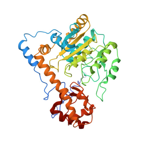
2.8-A-resolution crystal structure of an active-site mutant of aspartate aminotransferase from Escherichia coli.

Smith, D.L.Almo, S.C.Toney, M.D.Ringe, D.

(1989) Biochemistry 28: 8161-8167

  • DOI: https://doi.org/10.1021/bi00446a030
  • Primary Citation Related Structures: 
    2AAT

  • PubMed Abstract: 

    The three-dimensional structure of a mutant of the aspartate aminotransferase from Escherichia coli, in which the active-site lysine has been substituted by alanine (K258A), has been determined at 2.8-A resolution by X-ray diffraction. The mutant enzyme contains pyridoxamine phosphate as cofactor. The structure is compared to that of the mitochondrial aspartate aminotransferase. The most striking differences, aside from the absence of the lysine side chain, occur in the positions of the pyridoxamine group and of tryptophan 140.


  • Organizational Affiliation
    • Department of Chemistry, Massachusetts Institute of Technology, Cambridge 02139.

Macromolecule Content 

  • Total Structure Weight: 43.91 kDa 
  • Atom Count: 3,086 
  • Modeled Residue Count: 396 
  • Deposited Residue Count: 396 
  • Unique protein chains: 1

Macromolecules

Find similar proteins by:|  3D Structure
Entity ID: 1
MoleculeChains  Sequence LengthOrganismDetailsImage
ASPARTATE AMINOTRANSFERASE396Escherichia coliMutation(s): 1 
EC: 2.6.1.1
UniProt
Find proteins for P00509 (Escherichia coli (strain K12))
Explore P00509 
Go to UniProtKB:  P00509
Entity Groups
Sequence Clusters30% Identity50% Identity70% Identity90% Identity95% Identity100% Identity
UniProt GroupP00509
Sequence Annotations
Expand
Reference Sequence

Small Molecules

Ligands 2 Unique
IDChains Name / Formula / InChI Key2D Diagram3D Interactions
PMP

Query on PMP


Download:Ideal Coordinates CCD File
C [auth A]4'-DEOXY-4'-AMINOPYRIDOXAL-5'-PHOSPHATE
C8 H13 N2 O5 P
ZMJGSOSNSPKHNH-UHFFFAOYSA-N
SO4

Query on SO4


Download:Ideal Coordinates CCD File
B [auth A]SULFATE ION
O4 S
QAOWNCQODCNURD-UHFFFAOYSA-L

Experimental Data & Validation

Experimental Data

  • Method: X-RAY DIFFRACTION
  • Resolution: 2.80 Å
  • R-Value Observed: 0.220 (Depositor) 
Space Group: C 2 2 21
Unit Cell:
Length ( Å )Angle ( ˚ )
a = 156.8α = 90
b = 86.74β = 90
c = 79.84γ = 90
Software Package:
Software NamePurpose
PROLSQrefinement

Structure Validation

View Full Validation Report


Entry History 

Deposition Data

Revision History  (Full details and data files)

  • Version 1.0: 1989-10-15
    Type: Initial release
  • Version 1.1: 2008-03-24
    Changes: Version format compliance
  • Version 1.2: 2011-07-13
    Changes: Derived calculations, Version format compliance
  • Version 1.3: 2012-07-25
    Changes: Structure summary
  • Version 1.4: 2020-07-29
    Changes: Database references, Derived calculations, Other, Structure summary
  • Version 1.5: 2024-02-14
    Changes: Data collection, Database references