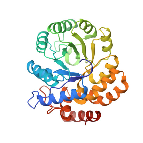
Role of Arginine 138 in the Catalysis and Regulation of Escherichia coli Dihydrodipicolinate Synthase.
Dobson, R.C., Devenish, S.R., Turner, L.A., Clifford, V.R., Pearce, F.G., Jameson, G.B., Gerrard, J.A.(2005) Biochemistry 44: 13007-13013
- PubMed: 16185069
- DOI: https://doi.org/10.1021/bi051281w
- Primary Citation of Related Structures:
2A6L, 2A6N - PubMed Abstract:
In plants and bacteria, the branch point of (S)-lysine biosynthesis is the condensation of (S)-aspartate-beta-semialdehyde [(S)-ASA] and pyruvate, a reaction catalyzed by dihydrodipicolinate synthase (DHDPS, EC 4.2.1.52). It has been proposed that Arg138, a residue situated at the entrance to the active site of DHDPS, is responsible for binding the carboxyl of (S)-ASA and may additionally be involved in the mechanism of (S)-lysine inhibition. This study tests these assertions by mutation of Arg138 to both histidine and alanine. Following purification, DHDPS-R138H and DHDPS-R138A each showed severely compromised activity (approximately 0.1% that of the wild type), and the apparent Michaelis-Menten constant for (S)-ASA in each mutant, calculated using a pseudo-single substrate analysis, was significantly higher than that of the wild type. This provides good evidence that Arg138 is indeed essential for catalysis and plays a key role in substrate binding. To test whether structural changes could account for the change in kinetic behavior, the solution structure was probed via far-UV circular dichroism, confirming that the mutations at position 138 did not modify secondary structure. The crystal structures of both mutant enzymes were determined, confirming the presence of the mutations and suggesting that Arg138 plays an important role in catalysis: the stabilization of the catalytic triad residues, a motif we have previously demonstrated to be essential for activity. In addition, the role of Arg138 in (S)-lysine inhibition was examined. Both mutant enzymes showed the same IC(50) values as the wild type but different partial inhibition patterns, from which it is concluded that arginine 138 is not essential for (S)-lysine inhibition.
- School of Biological Sciences, University of Canterbury, Private Bag 4800, Christchurch, New Zealand. renwick.dobson@canterbury.ac.nz
Organizational Affiliation:


